SCIENTIFIC COMPUTING AND IMAGING INSTITUTEat the University of Utah
An internationally recognized leader in visualization, scientific computing, and image analysis
SCI Publications
2014
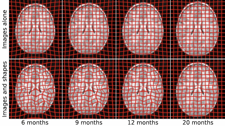
Geodesic Regression of Image and Shape Data for Improved Modeling of {4D} Trajectories
J. Fishbaugh, M. Prastawa, G. Gerig, S. Durrleman.
“Geodesic Regression of Image and Shape Data for Improved Modeling of 4D Trajectories,” In Proceedings of the 2014 IEEE International Symposium on Biomedical Imaging (ISBI), pp. (accepted). 2014.
ABSTRACT
×
A variety of regression schemes have been proposed on images or shapes, although available methods do not handle them jointly. In this paper, we present a framework for joint image and shape regression which incorporates images as well as anatomical shape information in a consistent manner. Evolution is described by a generative model that is the analog of linear regression, which is fully characterized by baseline images and shapes (intercept) and initial momenta vectors (slope). Further, our framework adopts a control point parameterization of deformations, where the dimensionality of the deformation is determined by the complexity of anatomical changes in time rather than the sampling of the image and/or the geometric data. We derive a gradient descent algorithm which simultaneously estimates baseline images and shapes, location of control points, and momenta. Experiments on real medical data demonstrate that our framework effectively combines image and shape information, resulting in improved modeling of 4D (3D space + time) trajectories.
A Joint Framework for {4D} Segmentation and Estimation of Smooth Temporal Appearance Changes
Y. Gao, M. Prastawa, M. Styner, J. Piven, G. Gerig.
“A Joint Framework for 4D Segmentation and Estimation of Smooth Temporal Appearance Changes,” In Proceedings of the 2014 IEEE International Symposium on Biomedical Imaging (ISBI), pp. (accepted). 2014.
ABSTRACT
×
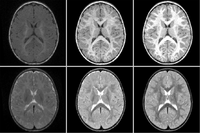
Medical imaging studies increasingly use longitudinal images of individual subjects in order to follow-up changes due to development, degeneration, disease progression or efficacy of therapeutic intervention. Repeated image data of individuals are highly correlated, and the strong causality of information over time lead to the development of procedures for joint segmentation of the series of scans, called 4D segmentation. A main aim was improved consistency of quantitative analysis, most often solved via patient-specific atlases. Challenging open problems are contrast changes and occurance of subclasses within tissue as observed in multimodal MRI of infant development, neurodegeneration and disease. This paper proposes a new 4D segmentation framework that enforces continuous dynamic changes of tissue contrast patterns over time as observed in such data. Moreover, our model includes the capability to segment different contrast patterns within a specific tissue class, for example as seen in myelinated and unmyelinated white matter regions in early brain development. Proof of concept is shown with validation on synthetic image data and with 4D segmentation of longitudinal, multimodal pediatric MRI taken at 6, 12 and 24 months of age, but the methodology is generic w.r.t. different application domains using serial imaging.
Prenatal cocaine effects on brain structure in early infancy
K. Grewen, M. Burchinal, C. Vachet, S. Gouttard, J.H. Gilmore, W. Lin, J. Johns, M. Elam, G. Gerig.
“Prenatal cocaine effects on brain structure in early infancy,” In NeuroImage, Vol. 101, pp. 114--123. November, 2014.
DOI: 10.1016/j.neuroimage.2014.06.070
ABSTRACT
×
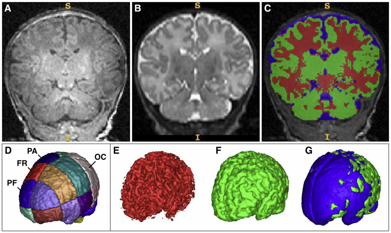
Prenatal cocaine exposure (PCE) is related to subtle deficits in cognitive and behavioral function in infancy, childhood and adolescence. Very little is known about the effects of in utero PCE on early brain development that may contribute to these impairments. The purpose of this study was to examine brain structural differences in infants with and without PCE. We conducted MRI scans of newborns (mean age = 5 weeks) to determine cocaine's impact on early brain structural development. Subjects were three groups of infants: 33 with PCE co-morbid with other drugs, 46 drug-free controls and 40 with prenatal exposure to other drugs (nicotine, alcohol, marijuana, opiates, SSRIs) but without cocaine. Infants with PCE exhibited lesser total gray matter (GM) volume and greater total cerebral spinal fluid (CSF) volume compared with controls and infants with non-cocaine drug exposure. Analysis of regional volumes revealed that whole brain GM differences were driven primarily by lesser GM in prefrontal and frontal brain regions in infants with PCE, while more posterior regions (parietal, occipital) did not differ across groups. Greater CSF volumes in PCE infants were present in prefrontal, frontal and parietal but not occipital regions. Greatest differences (GM reduction, CSF enlargement) in PCE infants were observed in dorsal prefrontal cortex. Results suggest that PCE is associated with structural deficits in neonatal cortical gray matter, specifically in prefrontal and frontal regions involved in executive function and inhibitory control. Longitudinal study is required to determine whether these early differences persist and contribute to deficits in cognitive functions and enhanced risk for drug abuse seen at school age and in later life.
Network inefficiencies in autism spectrum disorder at 24 months
J.D. Lewis, A.C. Evans, J.R. Pruett, K. Botteron, L. Zwaigenbaum, A. Estes, G. Gerig, L. Collins, P. Kostopoulos, R. McKinstry, S. Dager, S. Paterson, R. Schultz, M. Styner, H. Hazlett, J. Piven, IBIS network.
“Network inefficiencies in autism spectrum disorder at 24 months,” In Translational Psychiatry, Vol. 4, No. 5, Nature Publishing Group, pp. e388. May, 2014.
DOI: 10.1038/tp.2014.24
ABSTRACT
Autism Spectrum Disorder (ASD) is a developmental disorder defined by behavioural symptoms that emerge during the first years of life. Associated with these symptoms are differences in the structure of a wide array of brain regions, and in the connectivity between these regions. However, the use of cohorts with large age variability and participants past the generally recognized age of onset of the defining behaviours means that many of the reported abnormalities may be a result of cascade effects of developmentally earlier deviations. This study assessed differences in connectivity in ASD at the age at which the defining behaviours first become clear. The participants were 113 24-month-olds at high risk for ASD, 31 of whom were classified as ASD, and 23 typically developing 24-month-olds at low risk for ASD. Utilizing diffusion data to obtain measures of the length and strength of connections between anatomical regions, we performed an analysis of network efficiency. Our results showed significantly decreased local and global efficiency over temporal, parietal, and occipital lobes in high-risk infants classified as ASD, relative to both low- and high-risk infants not classified as ASD. The frontal lobes showed only a reduction in global efficiency in Broca's area. Additionally, these same regions showed an inverse relation between efficiency and symptom severity across the high-risk infants. The results suggest delay or deficits in infants with ASD in the optimization of both local and global aspects of network structure in regions involved in processing auditory and visual stimuli, language, and nonlinguistic social stimuli.
Diffeomorphic Shape Trajectories for Improved Longitudinal Segmentation and Statistics
P. Muralidharan, J. Fishbaugh, H.J. Johnson, S. Durrleman, J.S. Paulsen, G. Gerig, P.T. Fletcher.
“Diffeomorphic Shape Trajectories for Improved Longitudinal Segmentation and Statistics,” In Proceedings of Medical Image Computing and Computer Assisted Intervention (MICCAI), 2014.
ABSTRACT
×
Longitudinal imaging studies involve tracking changes in individuals by repeated image acquisition over time. The goal of these studies is to quantify biological shape variability within and across individuals, and also to distinguish between normal and disease populations. However, data variability is influenced by outside sources such as image acquisition, image calibration, human expert judgment, and limited robustness of segmentation and registration algorithms. In this paper, we propose a two-stage method for the statistical analysis of longitu- dinal shape. In the first stage, we estimate diffeomorphic shape trajectories for each individual that minimize inconsistencies in segmented shapes across time. This is followed by a longitudinal mixed-effects statistical model in the second stage for testing differences in shape trajectories between groups. We apply our method to a longitudinal database from PREDICT-HD and demonstrate our ap- proach reduces unwanted variability for both shape and derived measures, such as volume. This leads to greater statistical power to distinguish differences in shape trajectory between healthy subjects and subjects with a genetic biomarker for Huntington's disease (HD).
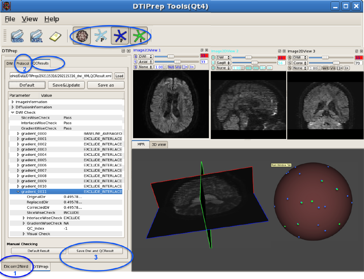
{DTIPrep:} Quality Control of Diffusion-Weighted Images
I. Oguz, M. Farzinfar, J. Matsui, F. Budin, Z. Liu, G. Gerig, H.J. Johnson, M.A. Styner.
“DTIPrep: Quality Control of Diffusion-Weighted Images,” In Frontiers in Neuroinformatics, Vol. 8, No. 4, 2014.
DOI: 10.3389/fninf.2014.00004
ABSTRACT
×
In the last decade, diffusion MRI (dMRI) studies of the human and animal brain have been used to investigate a multitude of pathologies and drug-related effects in neuroscience research. Study after study identifies white matter (WM) degeneration as a crucial biomarker for all these diseases. The tool of choice for studying WM is dMRI. However, dMRI has inherently low signal-to-noise ratio and its acquisition requires a relatively long scan time; in fact, the high loads required occasionally stress scanner hardware past the point of physical failure. As a result, many types of artifacts implicate the quality of diffusion imagery. Using these complex scans containing artifacts without quality control (QC) can result in considerable error and bias in the subsequent analysis, negatively affecting the results of research studies using them. However, dMRI QC remains an under-recognized issue in the dMRI community as there are no user-friendly tools commonly available to comprehensively address the issue of dMRI QC. As a result, current dMRI studies often perform a poor job at dMRI QC.
Thorough QC of diffusion MRI will reduce measurement noise and improve reproducibility, and sensitivity in neuroimaging studies; this will allow researchers to more fully exploit the power of the dMRI technique and will ultimately advance neuroscience. Therefore, in this manuscript, we present our open-source software, DTIPrep, as a unified, user friendly platform for thorough quality control of dMRI data. These include artifacts caused by eddy-currents, head motion, bed vibration and pulsation, venetian blind artifacts, as well as slice-wise and gradient-wise intensity inconsistencies. This paper summarizes a basic set of features of DTIPrep described earlier and focuses on newly added capabilities related to directional artifacts and bias analysis.
Semi-automated application for kidney motion correction and filtration analysis in {MR} renography
F. Rousset, C. Vachet, C. Conlin, M. Heilbrun, J.L. Zhang, V.S. Lee, G. Gerig.
“Semi-automated application for kidney motion correction and filtration analysis in MR renography,” In Proceeding of the 2014 Joint Annual Meeting ISMRM-ESMRMB, pp. (accepted). 2014.
ABSTRACT
Altered renal function commonly affects patients with cirrhosis, a consequence of chronic liver disease. From lowdose contrast material-enhanced magnetic resonance (MR) renography, we can estimate the Glomerular Filtration Rate (GFR), an important parameter to assess renal function. Two-dimensional MR images are acquired every 2 seconds for approximately 5 minutes during free breathing, which results in a dynamic series of 140 images representing kidney filtration over time. This specific acquisition presents dynamic contrast changes but is also challenged by organ motion due to breathing. Rather than use conventional image registration techniques, we opted for an alternative method based on object detection. We developed a novel analysis framework available under a stand-alone toolkit to efficiently register dynamic kidney series, manually select regions of interest, visualize the concentration curves for these ROIs, and fit them into a model to obtain GFR values. This open-source cross-platform application is written in C++, using the Insight Segmentation and Registration Toolkit (ITK) library, and QT4 as a graphical user interface.
Normative Modeling of Early Brain Maturation from Longitudinal {DTI} Reveals Twin-Singleton Differences
N. Sadeghi, J.H. Gilmore, W. Lin, G. Gerig.
“Normative Modeling of Early Brain Maturation from Longitudinal DTI Reveals Twin-Singleton Differences,” In Proceeding of the 2014 Joint Annual Meeting ISMRM-ESMRMB, pp. (accepted). 2014.
ABSTRACT
Early brain development of white matter is characterized by rapid organization and structuring. Magnetic Resonance diffusion tensor imaging (MR-DTI) provides the possibility of capturing these changes non-invasively by following individuals longitudinally in order to better understand departures from normal brain development in subjects at risk for mental illness [1]. Longitudinal imaging of individuals suggests the use of 4D (3D, time) image analysis and longitudinal statistical modeling [3].
Subject-specific prediction using nonlinear population modeling: Application to early brain maturation from {DTI}
N. Sadeghi, P.T. Fletcher, M. Prastawa, J.H. Gilmore, G. Gerig.
“Subject-specific prediction using nonlinear population modeling: Application to early brain maturation from DTI,” In Proceedings of Medical Image Computing and Computer-Assisted Intervention (MICCAI 2014), 2014.
ABSTRACT
×
The term prediction implies expected outcome in the future, often based on a model and statistical inference. Longitudinal imaging studies offer the possibility to model temporal change trajectories of anatomy across populations of subjects. In the spirit of subject-specific analysis, such normative models can then be used to compare data from new subjects to the norm and to study progression of disease or to predict outcome. This paper follows a statistical inference approach and presents a framework for prediction of future observations based on past measurements and population statistics. We describe prediction in the context of nonlinear mixed effects modeling (NLME) where the full reference population's statistics (estimated fixed effects, variance-covariance of random effects, variance of noise) is used along with the individual's available observations to predict its trajectory. The proposed methodology is generic in regard to application domains. Here, we demonstrate analysis of early infant brain maturation from longitudinal DTI with up to three time points. Growth as observed in DTI-derived scalar invariants is modeled with a parametric function, its parameters being input to NLME population modeling. Trajectories of new subject's data are estimated when using no observation, only the rst or the first two time points. Leave-one-out experiments result in statistics on differences between actual and predicted observations. We also simulate a clinical scenario of prediction on multiple categories, where trajectories predicted from multiple models are classified based on maximum likelihood criteria.
Parametric Regression Scheme for Distributions: Analysis of {DTI} Fiber Tract Diffusion Changes in Early Brain Development
A. Sharma, P.T. Fletcher, J.H. Gilmore, M.L. Escolar, A. Gupta, M. Styner, G. Gerig.
“Parametric Regression Scheme for Distributions: Analysis of DTI Fiber Tract Diffusion Changes in Early Brain Development,” In Proceedings of the 2014 IEEE International Symposium on Biomedical Imaging (ISBI), pp. (accepted). 2014.
ABSTRACT
×
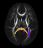
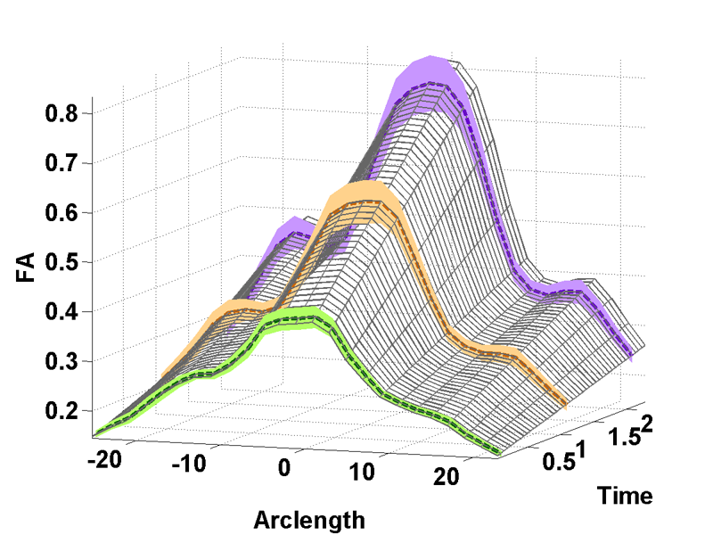
Temporal modeling frameworks often operate on scalar variables by summarizing data at initial stages as statistical summaries of the underlying distributions. For instance, DTI analysis often employs summary statistics, like mean, for regions of interest and properties along fiber tracts for population studies and hypothesis testing. This reduction via discarding of variability information may introduce significant errors which propagate through the procedures. We propose a novel framework which uses distribution-valued variables to retain and utilize the local variability information. Classic linear regression is adapted to employ these variables for model estimation. The increased stability and reliability of our proposed method when compared with regression using single-valued statistical summaries, is demonstrated in a validation experiment with synthetic data. Our driving application is the modeling of age-related changes along DTI white matter tracts. Results are shown for the spatiotemporal population trajectory of genu tract estimated from 45 healthy infants and compared with a Krabbe's patient.
Keywords: linear regression, distribution-valued data, spatiotemporal growth trajectory, DTI, early neurodevelopment
{4D} Modeling of Infant Brain Growth in Down's Syndrome and Controls from longitudinal {MRI}
C. Vachet, H.C. Hazlett, J. Piven, G. Gerig.
“4D Modeling of Infant Brain Growth in Down's Syndrome and Controls from longitudinal MRI,” In Proceeding of the 2014 Joint Annual Meeting ISMRM-ESMRMB, pp. (accepted). 2014.
ABSTRACT
Modeling of early brain growth trajectories from longitudinal MRI will provide new insight into neurodevelopmental characteristics, timing and type of changes in neurological disorders from controls. In addition to an ongoing large-scale infant autism neuroimaging study 1, we recruited 4 infants with Down’s syndrome (DS) in order to evaluate newly developed methods for 4D segmentation from longitudinal infant MRI, and for temporal modeling of brain growth trajectories. Specifically to Down's, a comparison of patterns of full brain and lobar tissue growth may lead to better insight into the observed variability of cognitive development and neurological effects, and may help with development of disease-modifying therapeutic intervention.
Characterizing growth patterns in longitudinal {MRI} using image contrast
A. Vardhan, M. Prastawa, C. Vachet, J. Piven, G. Gerig.
“Characterizing growth patterns in longitudinal MRI using image contrast,” In Proceedings of Medical Imaging 2014: Image Processing, 2014.
ABSTRACT
×
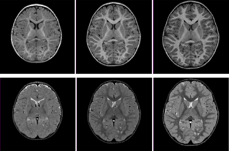
Understanding the growth patterns of the early brain is crucial to the study of neuro-development. In the early stages of brain growth, a rapid sequence of biophysical and chemical processes take place. A crucial component of these processes, known as myelination, consists of the formation of a myelin sheath around a nerve fiber, enabling the effective transmission of neural impulses. As the brain undergoes myelination, there is a subsequent change in the contrast between gray matter and white matter as observed in MR scans. In this work, graywhite matter contrast is proposed as an effective measure of appearance which is relatively invariant to location, scanner type, and scanning conditions. To validate this, contrast is computed over various cortical regions for an adult human phantom. MR (Magnetic Resonance) images of the phantom were repeatedly generated using different scanners, and at different locations. Contrast displays less variability over changing conditions of scan compared to intensity-based measures, demonstrating that it is less dependent than intensity on external factors. Additionally, contrast is used to analyze longitudinal MR scans of the early brain, belonging to healthy controls and Down's Syndrome (DS) patients. Kernel regression is used to model subject-specific trajectories of contrast changing with time. Trajectories of contrast changing with time, as well as time-based biomarkers extracted from contrast modeling, show large differences between groups. The preliminary applications of contrast based analysis indicate its future potential to reveal new information not covered by conventional volumetric or deformation-based analysis, particularly for distinguishing between normal and abnormal growth patterns.
Joint Longitudinal Modeling of Brain Appearance in Multimodal {MRI} for the Characterization of Early Brain Developmental Processes
A. Vardhan, N. Sadeghi, C. Vachet, J. Piven, G. Gerig.
“Joint Longitudinal Modeling of Brain Appearance in Multimodal MRI for the Characterization of Early Brain Developmental Processes,” In Spatiotemporal Image Analysis for Longitudinal and Time-Series Image Data (STIA'14) , LNCS. MICCAI'14, Springer Verlag, June, 2014.
ABSTRACT
×
Early brain maturational processes such as myelination manifest as changes in the relative appearance of white-gray matter tissue classes in MR images. Imaging modalities such as T1W (T1-Weighted) and T2W (T2-Weighted) MRI each display specific patterns of appearance change associated with distinct neurobiological components of these maturational processes. In this paper we present a framework to jointly model multimodal appearance changes across time for a longitudinal imaging dataset, resulting in quantitative assessment of the patterns of early brain maturation not yet available to clinicians. We measure appearance by quantifying contrast between white and gray matter in terms of the distance between their intensity distributions, a method demonstrated to be relatively stable to interscan variability. A multivariate nonlinear mixed effects (NLME) model is used for joint statistical modeling of this contrast measure across multiple imaging modalities. The multivariate NLME procedure considers correlations between modalities in addition to intra-modal variability. The parameters of the logistic growth function used in NLME modeling provide useful quantitative information about the timing and progression of contrast change in multimodal datasets. Inverted patterns of relative white-gray matter intensity gradient that are observable in T1W scans with respect to T2W scans are characterized by the SIR (Signal Intensity Ratio). The CONTDIR (Contrast Direction) which measures the direction of the gradient at each time point relative to that in the adult-like scan adds a directional attribute to contrast. The major contribution of this paper is a framework for joint multimodal temporal modeling of white-gray matter MRI contrast change and estimation of subject-specific and population growth trajectories. Results confirm qualitative descriptions of growth patterns in pediatric radiology studies and our new quantitative modeling scheme has the potential to advance understanding of variability of brain tissue maturation and to eventually differentiate normal from abnormal growth for early diagnosis of pathology.
UNC-Utah NA-MIC framework for DTI fiber tract analysis
A.R. Verde, F. Budin, J.-B. Berger, A. Gupta, M. Farzinfar, A. Kaiser, M. Ahn, H. Johnson, J. Matsui, H.C. Hazlett, A. Sharma, C. Goodlett, Y. Shi, S. Gouttard, C. Vachet, J. Piven, H. Zhu, G. Gerig, M. Styner.
“UNC-Utah NA-MIC framework for DTI fiber tract analysis,” In Frontiers in Neuroinformatics, Vol. 7, No. 51, January, 2014.
DOI: 10.3389/fninf.2013.00051
ABSTRACT
×
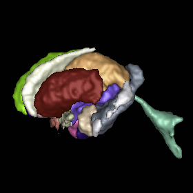
Diffusion tensor imaging has become an important modality in the field of neuroimaging to capture changes in micro-organization and to assess white matter integrity or development. While there exists a number of tractography toolsets, these usually lack tools for preprocessing or to analyze diffusion properties along the fiber tracts. Currently, the field is in critical need of a coherent end-to-end toolset for performing an along-fiber tract analysis, accessible to non-technical neuroimaging researchers. The UNC-Utah NA-MIC DTI framework represents a coherent, open source, end-to-end toolset for atlas fiber tract based DTI analysis encompassing DICOM data conversion, quality control, atlas building, fiber tractography, fiber parameterization, and statistical analysis of diffusion properties. Most steps utilize graphical user interfaces (GUI) to simplify interaction and provide an extensive DTI analysis framework for non-technical researchers/investigators. We illustrate the use of our framework on a small sample, cross sectional neuroimaging study of eight healthy 1-year-old children from the Infant Brain Imaging Study (IBIS) Network. In this limited test study, we illustrate the power of our method by quantifying the diffusion properties at 1 year of age on the genu and splenium fiber tracts.
Keywords: neonatal neuroimaging, white matter pathways, magnetic resonance imaging, diffusion tensor imaging, diffusion imaging quality control, DTI atlas building
{4D} Active Cut: An Interactive Tool for Pathological Anatomy Modeling
Bo Wang, W. Liu, M. Prastawa, A. Irimia, P.M. Vespa, J.D. van Horn, P.T. Fletcher, G. Gerig.
“4D Active Cut: An Interactive Tool for Pathological Anatomy Modeling,” In Proceedings of the 2014 IEEE International Symposium on Biomedical Imaging (ISBI), pp. (accepted). 2014.
ABSTRACT
×
4D pathological anatomy modeling is key to understanding complex pathological brain images. It is a challenging problem due to the difficulties in detecting multiple appearing and disappearing lesions across time points and estimating dynamic changes and deformations between them. We propose a novel semi-supervised method, called 4D active cut, for lesion recognition and deformation estimation. Existing interactive segmentation methods passively wait for user to refine the segmentations which is a difficult task in 3D images that change over time. 4D active cut instead actively selects candidate regions for querying the user, and obtains the most informative user feedback. A user simply answers 'yes' or 'no' to a candidate object without having to refine the segmentation slice by slice. Compared to single-object detection of the existing methods, our method also detects multiple lesions with spatial coherence using Markov random fields constraints. Results show improvement on the lesion detection, which subsequently improves deformation estimation.
Keywords: Active learning, graph cuts, longitudinal MRI, Markov Random Fields, semi-supervised learning
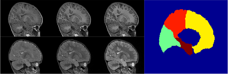
Multi-atlas segmentation of subcortical brain structures via the AutoSeg software pipeline
J. Wang, C. Vachet, A. Rumple, S. Gouttard, C. Ouzie, E. Perrot, G. Du, X. Huang, G. Gerig, M.A. Styner.
“Multi-atlas segmentation of subcortical brain structures via the AutoSeg software pipeline,” In Frontiers in Neuroinformatics, Vol. 8, No. 7, 2014.
DOI: 10.3389/fninf.2014.00007
ABSTRACT
×
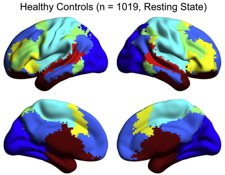
Automated segmenting and labeling of individual brain anatomical regions, in MRI are challenging, due to the issue of individual structural variability. Although atlas-based segmentation has shown its potential for both tissue and structure segmentation, due to the inherent natural variability as well as disease-related changes in MR appearance, a single atlas image is often inappropriate to represent the full population of datasets processed in a given neuroimaging study. As an alternative for the case of single atlas segmentation, the use of multiple atlases alongside label fusion techniques has been introduced using a set of individual “atlases” that encompasses the expected variability in the studied population. In our study, we proposed a multi-atlas segmentation scheme with a novel graph-based atlas selection technique. We first paired and co-registered all atlases and the subject MR scans. A directed graph with edge weights based on intensity and shape similarity between all MR scans is then computed. The set of neighboring templates is selected via clustering of the graph. Finally, weighted majority voting is employed to create the final segmentation over the selected atlases. This multi-atlas segmentation scheme is used to extend a single-atlas-based segmentation toolkit entitled AutoSeg, which is an open-source, extensible C++ based software pipeline employing BatchMake for its pipeline scripting, developed at the Neuro Image Research and Analysis Laboratories of the University of North Carolina at Chapel Hill. AutoSeg performs N4 intensity inhomogeneity correction, rigid registration to a common template space, automated brain tissue classification based skull-stripping, and the multi-atlas segmentation. The multi-atlas-based AutoSeg has been evaluated on subcortical structure segmentation with a testing dataset of 20 adult brain MRI scans and 15 atlas MRI scans. The AutoSeg achieved mean Dice coefficients of 81.73% for the subcortical structures.
Down Syndrome is the most common genetic cause for intellectual disability, yet the pathophysiology of cognitive impairment in Down Syndrome is unknown. We compared fMRI scans of 15 individuals with Down Syndrome to 14 typically developing control subjects while they viewed 50 min of cartoon video clips. There was widespread increased synchrony between brain regions, with only a small subset of strong, distant connections showing underconnectivity in Down Syndrome. Brain regions showing negative correlations were less anticorrelated and were among the most strongly affected connections in the brain. Increased correlation was observed between all of the distributed brain networks studied, with the strongest internetwork correlation in subjects with the lowest performance IQ. A functional parcellation of the brain showed simplified network structure in Down Syndrome organized by local connectivity. Despite increased interregional synchrony, intersubject correlation to the cartoon stimuli was lower in Down Syndrome, indicating that increased synchrony had a temporal pattern that was not in response to environmental stimuli, but idiosyncratic to each Down Syndrome subject. Short-range, increased synchrony was not observed in a comparison sample of 447 autism vs. 517 control subjects from the Autism Brain Imaging Exchange (ABIDE) collection of resting state fMRI data, and increased internetwork synchrony was only observed between the default mode and attentional networks in autism. These findings suggest immature development of connectivity in Down Syndrome with impaired ability to integrate information from distant brain regions into coherent distributed networks.
Toward a comprehensive framework for the spatiotemporal statistical analysis of longitudinal shape data
S. Durrleman, X. Pennec, A. Trouvé, J. Braga, G. Gerig, N. Ayache.
“Toward a comprehensive framework for the spatiotemporal statistical analysis of longitudinal shape data,” In International Journal of Computer Vision (IJCV), Vol. 103, No. 1, pp. 22--59. September, 2013.
DOI: 10.1007/s11263-012-0592-x
ABSTRACT
×
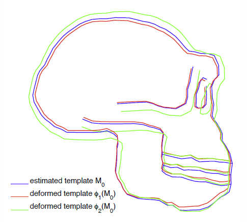
This paper proposes an original approach for the statistical analysis of longitudinal shape data. The proposed method allows the characterization of typical growth patterns and subject-specific shape changes in repeated time-series observations of several subjects. This can be seen as the extension of usual longitudinal statistics of scalar measurements to high-dimensional shape or image data.
The method is based on the estimation of continuous subject-specific growth trajectories and the comparison of such temporal shape changes across subjects. Differences between growth trajectories are decomposed into morphological deformations, which account for shape changes independent of the time, and time warps, which account for different rates of shape changes over time.
Given a longitudinal shape data set, we estimate a mean growth scenario representative of the population, and the variations of this scenario both in terms of shape changes and in terms of change in growth speed. Then, intrinsic statistics are derived in the space of spatiotemporal deformations, which characterize the typical variations in shape and in growth speed within the studied population. They can be used to detect systematic developmental delays across subjects.
In the context of neuroscience, we apply this method to analyze the differences in the growth of the hippocampus in children diagnosed with autism, developmental delays and in controls. Result suggest that group differences may be better characterized by a different speed of maturation rather than shape differences at a given age. In the context of anthropology, we assess the differences in the typical growth of the endocranium between chimpanzees and bonobos. We take advantage of this study to show the robustness of the method with respect to change of parameters and perturbation of the age estimates.
Frontolimbic neural circuitry at 6 months predicts individual differences in joint attention at 9 months
J.T. Elison, J.J. Wolff, D.C. Heimer, S.J. Paterson, H. Gu, M. Styner, G. Gerig, J. Piven, the IBIS Network.
“Frontolimbic neural circuitry at 6 months predicts individual differences in joint attention at 9 months,” In Developmental Science, Vol. 16, No. 2, Wiley-Blackwell, pp. 186--197. 2013.
DOI: 10.1111/desc.12015
PubMed Central ID: PMC3582040
ABSTRACT
×
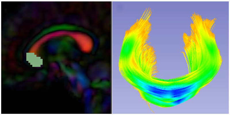
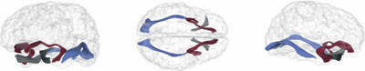
Elucidating the neural basis of joint attention in infancy promises to yield important insights into the development of language and social cognition, and directly informs developmental models of autism.We describe a new method for evaluating responding to joint attention performance in infancy that highlights the 9- to 10-month period as a time interval of maximal individual differences.We then demonstrate that fractional anisotropy in the right uncinate fasciculus, a white matter fiber bundle connecting the amygdala to the ventral-medial prefrontal cortex and anterior temporal pole, measured in 6-month-olds predicts individual differences in responding to joint attention at 9 months of age. The white matter microstructure of the right uncinate was not related to receptive language ability at 9 months. These findings suggest that the development of core nonverbal social communication skills in infancy is largely supported by preceding developments within right lateralized frontotemporal brain systems.
White Matter Microstructure and Atypical Visual Orienting in 7 Month-Olds at Risk for Autism
J.T. Elison, S.J. Paterson, J.J. Wolff, J.S. Reznick, N.J. Sasson, H. Gu, K.N. Botteron, S.R. Dager, A.M. Estes, A.C. Evans, G. Gerig, H.C. Hazlett, R.T. Schultz, M. Styner, L. Zwaigenbaum, J. Piven for the IBIS Network.
“White Matter Microstructure and Atypical Visual Orienting in 7 Month-Olds at Risk for Autism,” In American Journal of Psychiatry, Vol. AJP-12-09-1150.R2, March, 2013.
DOI: 10.1176/appi.ajp.2012.12091150
PubMed ID: 23511344
ABSTRACT
Objective: To determine whether specific patterns of oculomotor functioning and visual orienting characterize 7 month-old infants later classified with an autism spectrum disorder (ASD) and to identify the neural correlates of these behaviors.
Method: Ninety-seven infants contributed data to the current study (16 high-familial risk infants later classified with an ASD, 40 high-familial risk infants not meeting ASD criteria (high-risk-negative), and 41 low-risk infants). All infants completed an eye tracking task at 7 months and a clinical assessment at 25 months; diffusion weighted imaging data was acquired on 84 infants at 7 months. Primary outcome measures included average saccadic reaction time in a visually guided saccade procedure and radial diffusivity (an index of white matter organization) in fiber tracts that included corticospinal pathways and the splenium and genu of the corpus callosum.
Results: Visual orienting latencies were increased in seven-month-old infants who later express ASD symptoms at 25 months when compared with both high-risk-negative infants (p = 0.012, d = 0.73) and low-risk infants (p = 0.032, d = 0.71). Visual orienting latencies were uniquely associated with the microstructural organization of the splenium of the corpus callosum in low-risk infants, but this association was not apparent in infants later classified with ASD.
Conclusions: Flexibly and efficiently orienting to salient information in the environment is critical for subsequent cognitive and social-cognitive development. Atypical visual orienting may represent an earlyemerging prodromal feature of ASD, and abnormal functional specialization of posterior cortical circuits directly informs a novel model of ASD pathogenesis.