|
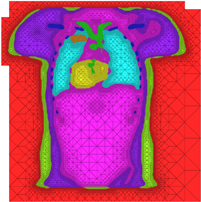
| A cross-section of a 3-dimensional, tetrahedral mesh of a torso. Each separate organ type is shown using a different color. |
This year, an essential goal has been to enhance the generalized image-processing pipeline of software developed by CIBC and its partners. With the growing use of high quality medical imaging, practitioners around the globe are employing these acquired datasets for performing biomedical simulation. In its holistic approach to image-to-simulation pipelines, our software starts with image data and processing, constructs geometric models, performs simulation, and provides biophysical analysis of the data. A research highlight for the CIBC this year is the development of an improved scheme for mesh generation, a critical step within this pipeline. This research complements the software package BioMesh3D, but targets a completely different niche in the world of mesh generation algorithms.
High Quality Meshing
The problem of mesh generation has been widely studied, as a hybrid field of interest to the scientific, engineering, and computer science communities. In each of these fields, meshes are used to compute numerical approximations to solutions of partial differential equations. To do so, continuous mathematics are replaced with a discrete analogue, most commonly to facilitate the finite element method (FEM).
The FEM works by decomposing a domain of interest into discrete entities of various dimensions, such as points (0-dimensional), edges (1-dimensional), and cells of higher dimension (frequently triangles and quadrilaterals are used for 2-dimensionl elements, tetrahedra and hexahedra for 3-dimensional). Together, these elements form what is commonly called a mesh (see figure)Solutions to the complex system are solved piecewise on each element, and then aggregated together to form the final solution. The FEM has become an important tool in medical imaging as well. For example, CT scans of legs can be meshed so that orthopedic modeling can accurately simulate gait, MRI scans of the torso are frequently used in cardiac electrophysical modeling, and images of the skull can identify structures of the brain.
Because the FEM is a computational tool that processes individual elements to approximate a whole solution, it is deeply impacted by the mesh elements used to represent the space. Two principle concerns stand out in the meshing problem for medical images: