 |
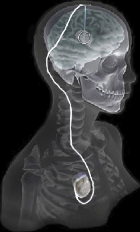
| Overview of the DBS system. The DBS electrode is implanted in the brain during stereotactic surgery. The electrode is attached via an extension wire to the IPG, which is implanted in the torso. The entire system is subcutaneous and is designed to deliver continuous stimulation for several years at a time. |
In recent years, there has been significant growth in the use of patient-specific models to predict the effects of neuromodulation therapies, such as deep brain stimulation (DBS). However, translating these models from a research environment to the everyday clinical workflow is a challenge, primarily due to the complexity of the models and the expertise required in specialized visualization software. Recently, the CIBC has worked with Dr. Christopher Butson at the University of Wisconsin to deploy the interactive visualization system ImageVis3D Mobile for experimental use in the area of DBS planning. In addition to running on multi-node compute clusters and large desktop systems, ImageVis3D is also designed for mobile computing devices such as the iPhone or iPad. In this case, ImageVis3D was modified for an evaluation environment in order to visualize models of Parkinson's Disease (PD) patients who received DBS therapy
1.
The selection of DBS settings is a significant clinical challenge that requires repeated revisions to achieve optimal therapeutic response, and is often performed without any visual representation of the stimulation system in the patient. We used ImageVis3D Mobile to provide models to movement disorders clinicians and asked them to use the software to determine: 1) which of the four DBS electrode contacts they would select for therapy and 2) what stimulation settings they would choose. We compared the stimulation protocol chosen from the software versus the stimulation protocol that was chosen via clinical practice (independent of the study). Lastly, we compared the amount of time required to reach these settings using the software versus the time required through standard practice. We found that the stimulation settings chosen using ImageVis3D Mobile were similar to those used in standard care, but were selected in drastically less time. We found that our visualization system, available directly at the point of care on a device familiar to the clinician, can be used to guide clinical decision-making for selecting DBS settings. The positive impact of the system could also translate to areas other than DBS.