
SCI Publications
2017
A. Vardhan, J. Fishbaugh, C. Vachet, G. Gerig.
“Longitudinal Modeling of Multi-modal Image Contrast Reveals Patterns of Early Brain Growth,” In Medical Image Computing and Computer Assisted Intervention - MICCAI 2017, Springer International Publishing, pp. 75--83. 2017.
DOI: 10.1007/978-3-319-66182-7_9
The brain undergoes rapid development during early childhood as a series of biophysical and chemical processes occur, which can be observed in magnetic resonance (MR) images as a change over time of white matter intensity relative to gray matter. Such a contrast change manifests in specific patterns in different imaging modalities, suggesting that brain maturation is encoded by appearance changes in multi-modal MRI. In this paper, we explore the patterns of early brain growth encoded by multi-modal contrast changes in a longitudinal study of children. For a given modality, contrast is measured by comparing histograms of intensity distributions between white and gray matter. Multivariate non-linear mixed effects (NLME) modeling provides subject-specific as well as population growth trajectories which accounts for contrast from multiple modalities. The multivariate NLME procedure and resulting non-linear contrast functions enable the study of maturation in various regions of interest. Our analysis of several brain regions in a study of 70 healthy children reveals a posterior to anterior pattern of timing of maturation in the major lobes of the cerebral cortex, with posterior regions maturing earlier than anterior regions. Furthermore, we find significant differences between maturation rates between males and females.
![]()
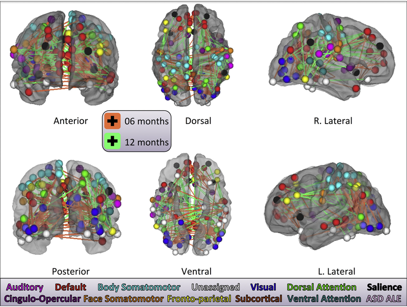
J. J. Wolff, M. R. Swanson, J. T. Elison, G. Gerig, J. R. Pruett, M. A. Styner, C. Vachet, K. N. Botteron, S. R. Dager, A. M. Estes, H. C. Hazlett, R. T. Schultz, M. D. Shen, L. Zwaigenbaum, J. Piven.
“Neural circuitry at age 6~months associated with later repetitive behavior and sensory responsiveness in autism,” In Molecular Autism, Vol. 8, No. 1, Springer Nature, March, 2017.
DOI: 10.1186/s13229-017-0126-z
Background
Restricted and repetitive behaviors are defining features of autism spectrum disorder (ASD). Under revised diagnostic criteria for ASD, this behavioral domain now includes atypical responses to sensory stimuli. To date, little is known about the neural circuitry underlying these features of ASD early in life.
Methods
Longitudinal diffusion tensor imaging data were collected from 217 infants at high familial risk for ASD. Forty-four of these infants were diagnosed with ASD at age 2. Targeted cortical, cerebellar, and striatal white matter pathways were defined and measured at ages 6, 12, and 24 months. Dependent variables included the Repetitive Behavior Scale-Revised and the Sensory Experiences Questionnaire.
Results
Among children diagnosed with ASD, repetitive behaviors and sensory response patterns were strongly correlated, even when accounting for developmental level or social impairment. Longitudinal analyses indicated that the genu and cerebellar pathways were significantly associated with both repetitive behaviors and sensory responsiveness but not social deficits. At age 6 months, fractional anisotropy in the genu significantly predicted repetitive behaviors and sensory responsiveness at age 2. Cerebellar pathways significantly predicted later sensory responsiveness. Exploratory analyses suggested a possible disordinal interaction based on diagnostic status for the association between fractional anisotropy and repetitive behavior.
Conclusions
Our findings suggest that restricted and repetitive behaviors contributing to a diagnosis of ASD at age 2 years are associated with structural properties of callosal and cerebellar white matter pathways measured during infancy and toddlerhood. We further identified that repetitive behaviors and unusual sensory response patterns co-occur and share common brain-behavior relationships. These results were strikingly specific given the absence of association between targeted pathways and social deficits.
2016
![]()
S. Elhabian, C. Vachet, J. Piven, M. Styner, G. Gerig.
“Compressive sensing based Q-space resampling for handling fast bulk motion in hardi acquisitions,” In 2016 IEEE 13th International Symposium on Biomedical Imaging (ISBI), IEEE, pp. 907--910. April, 2016.
DOI: 10.1109/isbi.2016.7493412
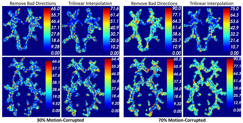
Diffusion-weighted (DW) MRI has become a widely adopted imaging modality to reveal the underlying brain connectivity. Long acquisition times and/or non-cooperative patients increase the chances of motion-related artifacts. Whereas slow bulk motion results in inter-gradient misalignment which can be handled via retrospective motion correction algorithms, fast bulk motion usually affects data during the application of a single diffusion gradient causing signal dropout artifacts. Common practices opt to discard gradients bearing signal attenuation due to the difficulty of their retrospective correction, with the disadvantage to lose full gradients for further processing. Nonetheless, such attenuation might only affect limited number of slices within a gradient volume. Q-space resampling has recently been proposed to recover corrupted slices while saving gradients for subsequent reconstruction. However, few corrupted gradients are implicitly assumed which might not hold in case of scanning unsedated infants or patients in pain. In this paper, we propose to adopt recent advances in compressive sensing based reconstruction of the diffusion orientation distribution functions (ODF) with under sampled measurements to resample corrupted slices. We make use of Simple Harmonic Oscillator based Reconstruction and Estimation (SHORE) basis functions which can analytically model ODF from arbitrary sampled signals. We demonstrate the impact of the proposed resampling strategy compared to state-of-art resampling and gradient exclusion on simulated intra-gradient motion as well as samples from real DWI data.
![]()
Y. Gao, M. Zhang, K. Grewen, P. T. Fletcher, G. Gerig.
“Image registration and segmentation in longitudinal MRI using temporal appearance modeling,” In 2016 IEEE 13th International Symposium on Biomedical Imaging (ISBI), IEEE, pp. 629--632. April, 2016.
DOI: 10.1109/isbi.2016.7493346
![]()
S. Kim, I.Lyu, V. Fonov, C. Vachet, H. Hazlett, R. Smith, J. Piven, S. Dager, R. Mckinstry, J. Pruett, A. Evans, D. Collins, K. Botteron, R. Schultz, G. Gerig, M. Styner.
“Development of Cortical Shape in the Human Brain from 6 to 24 Months of Age via a Novel Measure of Shape Complexity,” In NeuroImage, Vol. 135, Elsevier, pp. 163--176. July, 2016.
DOI: 10.1016/j.neuroimage.2016.04.053
The quantification of local surface morphology in the human cortex is important for examining population differences as well as developmental changes in neurodegenerative or neurodevelopmental disorders. We propose a novel cortical shape measure, referred to as the 'shape complexity index' (SCI), that represents localized shape complexity as the difference between the observed distributions of local surface topology, as quantified by the shape index (SI) measure, to its best fitting simple topological model within a given neighborhood. We apply a relatively small, adaptive geodesic kernel to calculate the SCI. Due to the small size of the kernel, the proposed SCI measure captures fine differences of cortical shape. With this novel cortical feature, we aim to capture comparatively small local surface changes that capture a) the widening versus deepening of sulcal and gyral regions, as well as b) the emergence and development of secondary and tertiary sulci. Current cortical shape measures, such as the gyrification index (GI) or intrinsic curvature measures, investigate the cortical surface at a different scale and are less well suited to capture these particular cortical surface changes. In our experiments, the proposed SCI demonstrates higher complexity in the gyral/sulcal wall regions, lower complexity in wider gyral ridges and lowest complexity in wider sulcal fundus regions. In early postnatal brain development, our experiments show that SCI reveals a pattern of increased cortical shape complexity with age, as well as sexual dimorphisms in the insula, middle cingulate, parieto-occipital sulcal and Broca's regions. Overall, sex differences were greatest at 6months of age and were reduced at 24months, with the difference pattern switching from higher complexity in males at 6months to higher complexity in females at 24months. This is the first study of longitudinal, cortical complexity maturation and sex differences, in the early postnatal period from 6 to 24months of age with fine scale, cortical shape measures. These results provide information that complement previous studies of gyrification index in early brain development.
![]()
P. Muralidharan, J. Fishbaugh, E. Y. Kim, H. J. Johnson, J. S. Paulsen, G. Gerig, P. T. Fletcher.
“Bayesian Covariate Selection in Mixed Effects Models for Longitudinal Shape Analysis,” In International Symposium on Biomedical Imaging (ISBI), IEEE, April, 2016.
DOI: 10.1109/isbi.2016.7493352
The goal of longitudinal shape analysis is to understand how anatomical shape changes over time, in response to biological processes, including growth, aging, or disease. In many imaging studies, it is also critical to understand how these shape changes are affected by other factors, such as sex, disease diagnosis, IQ, etc. Current approaches to longitudinal shape analysis have focused on modeling age-related shape changes, but have not included the ability to handle covariates. In this paper, we present a novel Bayesian mixed-effects shape model that incorporates simultaneous relationships between longitudinal shape data and multiple predictors or covariates to the model. Moreover, we place an Automatic Relevance Determination (ARD) prior on the parameters, that lets us automatically select which covariates are most relevant to the model based on observed data. We evaluate our proposed model and inference procedure on a longitudinal study of Huntington's disease from PREDICT-HD. We first show the utility of the ARD prior for model selection in a univariate modeling of striatal volume, and next we apply the full high-dimensional longitudinal shape model to putamen shapes.
2015
C.C. Conlin, J.L. Zhang, F. Rousset, C. Vachet, Y. Zhao, K.A. Morton, K. Carlston, G. Gerig, V.S. Lee.
“Performance of an Efficient Image-registration Algorithm in Processing MR Renography Data,” In J Magnetic Resonance Imaging, July, 2015.
DOI: 10.1002/jmri.25000
PURPOSE:
To evaluate the performance of an edge-based registration technique in correcting for respiratory motion artifacts in magnetic resonance renographic (MRR) data and to examine the efficiency of a semiautomatic software package in processing renographic data from a cohort of clinical patients.
MATERIALS AND METHODS:
The developed software incorporates an image-registration algorithm based on the generalized Hough transform of edge maps. It was used to estimate glomerular filtration rate (GFR), renal plasma flow (RPF), and mean transit time (MTT) from 36 patients who underwent free-breathing MRR at 3T using saturation-recovery turbo-FLASH. The processing time required for each patient was recorded. Renal parameter estimates and model-fitting residues from the software were compared to those from a previously reported technique. Interreader variability in the software was quantified by the standard deviation of parameter estimates among three readers. GFR estimates from our software were also compared to a reference standard from nuclear medicine.
RESULTS:
The time taken to process one patient's data with the software averaged 12 ± 4 minutes. The applied image registration effectively reduced motion artifacts in dynamic images by providing renal tracer-retention curves with significantly smaller fitting residues (P < 0.01) than unregistered data or data registered by the previously reported technique. Interreader variability was less than 10% for all parameters. GFR estimates from the proposed method showed greater concordance with reference values (P < 0.05).
CONCLUSION:
These results suggest that the proposed software can process MRR data efficiently and accurately. Its incorporated registration technique based on the generalized Hough transform effectively reduces respiratory motion artifacts in free-breathing renographic acquisitions. J. Magn. Reson. Imaging 2015.
S. Durrleman, T.P. Fletcher, G. Gerig, M. Niethammer, X. Pennec (Eds.).
“Spatio-temporal Image Analysis for Longitudinal and Time-Series Image Data,” In Proceedings of the Third International Workshop, STIA 2014, Image Processing, Computer Vision, Pattern Recognition, and Graphics, Vol. 8682, Springer LNCS, 2015.
ISBN: 978-3-319-14905-9
This book constitutes the thoroughly refereed post-conference proceedings of the Third
International Workshop on Spatio-temporal Image Analysis for Longitudinal and Time-
Series Image Data, STIA 2014, held in conjunction with MICCAI 2014 in Boston, MA, USA, in
September 2014.
The 7 papers presented in this volume were carefully reviewed and selected from 15
submissions. They are organized in topical sections named: longitudinal registration and
shape modeling, longitudinal modeling, reconstruction from longitudinal data, and 4D
image processing.
J.R. Pruett Jr., S. Kandala, S. Hoertel, A.Z. Snyder, J.T. Elison, T. Nishino, E. Feczko, N.U.F. Dosenbach, B. Nardos, J.D. Power, B. Adeyemo, K.N. Botteron, R.C. McKinstry, A.C. Evans, H.C. Hazlett, S.R. Dager, S. Paterson, R.T. Schultz, D.L. Collins, V.S. Fonov, M. Styner, G. Gerig, S. Das, P. Kostopoulos, J.N. Constantino, A.M. Estes, The IBIS Network, S.E. Petersen, B.L. Schlaggar, J. Piven.
“Accurate age classification of 6 and 12 month-old infants based on resting-state functional connectivity magnetic resonance imaging data,” In Developmental Cognitive Neuroscience, Vol. 12, pp. 123--133. April, 2015.
DOI: 10.1016/j.dcn.2015.01.003
![]()
S. Pujol, W. Wells, C. Pierpaoli, C. Brun, J. Gee, G. Cheng, B. Vemuri, O. Commowick, S. Prima, A. Stamm, M. Goubran, A. Khan, T. Peters, P. Neher, K. H. Maier-Hein, Y. Shi, A. Tristan-Vega, G. Veni, R. Whitaker, M. Styner, C.F. Westin, S. Gouttard, I. Norton, L. Chauvin, H. Mamata, G. Gerig, A. Nabavi, A. Golby,, R. Kikinis.
“The DTI Challenge: Toward Standardized Evaluation of Diffusion Tensor Imaging Tractography for Neurosurgery,” In Journal of Neuroimaging, Wiley, August, 2015.
DOI: 10.1111/jon.12283

Diffusion tensor imaging (DTI) tractography reconstruction of white matter pathways can help guide brain tumor resection. However, DTI tracts are complex mathematical objects and the validity of tractography-derived information in clinical settings has yet to be fully established. To address this issue, we initiated the DTI Challenge, an international working group of clinicians and scientists whose goal was to provide standardized evaluation of tractography methods for neurosurgery. The purpose of this empirical study was to evaluate different tractography techniques in the first DTI Challenge workshop.
METHODS
Eight international teams from leading institutions reconstructed the pyramidal tract in four neurosurgical cases presenting with a glioma near the motor cortex. Tractography methods included deterministic, probabilistic, filtered, and global approaches. Standardized evaluation of the tracts consisted in the qualitative review of the pyramidal pathways by a panel of neurosurgeons and DTI experts and the quantitative evaluation of the degree of agreement among methods.
RESULTS
The evaluation of tractography reconstructions showed a great interalgorithm variability. Although most methods found projections of the pyramidal tract from the medial portion of the motor strip, only a few algorithms could trace the lateral projections from the hand, face, and tongue area. In addition, the structure of disagreement among methods was similar across hemispheres despite the anatomical distortions caused by pathological tissues.
CONCLUSIONS
The DTI Challenge provides a benchmark for the standardized evaluation of tractography methods on neurosurgical data. This study suggests that there are still limitations to the clinical use of tractography for neurosurgical decision making.
![]()
H. J.V. Rutherford, G. Gerig, S. Gouttard, M. N. Potenza, L. C. Mayes.
“Investigating maternal brain structure and its relationship to substance use and motivational systems,” In Yale Journal of Biology and Medicine, in print, 2015.
Substance use during pregnancy and the postpartum period may have significant implications for both mother and the developing child. However, the neurobiological basis of the impact of substance use on parenting is less well understood. Here we examined the impact of maternal substance use on cortical gray matter (GM) and white matter volumes, and whether this was associated with individual differences in motivational systems of behavioral activation and inhibition. Mothers were included in the substance-using group if any addictive substance was used during pregnancy and/or in the immediate postpartum period (within 3 months of delivery). GM volume was reduced in substance-using mothers compared to non-substance-using mothers, particularly in frontal brain regions. In substance-using mothers, we also found that frontal GM was negatively correlated with levels of behavioral activation (i.e., the motivation to approach rewarding stimuli). This effect was absent in non-substance-using mothers. Taken together, these findings indicate a reduction in GM volume is associated with substance use, and that frontal GM volumetric differences may be related to approach motivation in substance-using mothers.
![]()
N. Sadeghi, J. H. Gilmore , G. Gerig.
“Modeling Brain Growth and Development,” In Brain, Vol. 1, pp. 429-436. 2015.
DOI: 10.1016/B978-0-12-397025-1.00314-6
![]()
A. P. Salzwedel, K. M. Grewen, C. Vachet, G. Gerig, W. Lin,, W. Gao.
“Prenatal Drug Exposure Affects Neonatal Brain Functional Connectivity,” In The Journal of Neuroscience, Vol. 35, No. 14, pp. 5860-5869. April, 2015.
DOI: 10.1523/JNEUROSCI.4333-14.2015
![]()
M. R. Swanson, J. J. Wolff, J. T. Elison, H. Gu, H. C. Hazlett, K. Botteron, M. Styner, S. Paterson, G. Gerig, J. Constantino, S. Dager, A. Estes, C. Vachet, J. Piven.
“Splenium development and early spoken language in human infants,” In Developmental Science, Wiley Online Library, 2015.
ISSN: 1467-7687
DOI: 10.1111/desc.12360
The association between developmental trajectories of language-related white matter fiber pathways from 6 to 24 months of age and individual differences in language production at 24 months of age was investigated. The splenium of the corpus callosum, a fiber pathway projecting through the posterior hub of the default mode network to occipital visual areas, was examined as well as pathways implicated in language function in the mature brain, including the arcuate fasciculi, uncinate fasciculi, and inferior longitudinal fasciculi. The hypothesis that the development of neural circuitry supporting domain-general orienting skills would relate to later language performance was tested in a large sample of typically developing infants. The present study included 77 infants with diffusion weighted MRI scans at 6, 12 and 24 months and language assessment at 24 months. The rate of change in splenium development varied significantly as a function of language production, such that children with greater change in fractional anisotropy (FA) from 6 to 24 months produced more words at 24 months. Contrary to findings from older children and adults, significant associations between language production and FA in the arcuate, uncinate, or left inferior longitudinal fasciculi were not observed. The current study highlights the importance of tracing brain development trajectories from infancy to fully elucidate emerging brain–behavior associations while also emphasizing the role of the splenium as a key node in the structural network that supports the acquisition of spoken language.
![]()
J. J. Wolff, G. Gerig, J. D. Lewis, T. Soda, M. A. Styner, C. Vachet, K. N. Botteron, J. T. Elison, S. R. Dager, A. M. Estes, H. C. Hazlett, R. T. Schultz, L. Zwaigenbaum, J. Piven.
“Altered corpus callosum morphology associated with autism over the first 2 years of life,” In Brain, 2015.
DOI: 10.1093/brain/awv118

2014
![]()
S. Durrleman, M. Prastawa, N. Charon, J.R. Korenberg, S. Joshi, G. Gerig, A. Trouvé.
“Morphometry of anatomical shape complexes with dense deformations and sparse parameters,” In NeuroImage, 2014.
DOI: 10.1016/j.neuroimage.2014.06.043
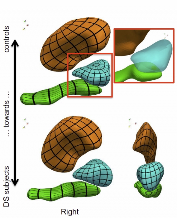
We propose a generic method for the statistical analysis of collections of anatomical shape complexes, namely sets of surfaces that were previously segmented and labeled in a group of subjects. The method estimates an anatomical model, the template complex, that is representative of the population under study. Its shape reflects anatomical invariants within the dataset. In addition, the method automatically places control points near the most variable parts of the template complex. Vectors attached to these points are parameters of deformations of the ambient 3D space. These deformations warp the template to each subject’s complex in a way that preserves the organization of the anatomical structures. Multivariate statistical analysis is applied to these deformation parameters to test for group differences. Results of the statistical analysis are then expressed in terms of deformation patterns of the template complex, and can be visualized and interpreted.
The user needs only to specify the topology of the template complex and the number of control points. The method then automatically estimates the shape of the template complex, the optimal position of control points and deformation parameters. The proposed approach is completely generic with respect to any type of application and well adapted to efficient use in clinical studies, in that it does not require point correspondence across surfaces and is robust to mesh imperfections such as holes, spikes, inconsistent orientation or irregular meshing.
The approach is illustrated with a neuroimaging study of Down syndrome (DS). Results demonstrate that the complex of deep brain structures shows a statistically significant shape difference between control and DS subjects. The deformation-based modelingis able to classify subjects with very high specificity and sensitivity, thus showing important generalization capability even given a low sample size. We show that results remain significant even if the number of control points, and hence the dimension of variables in the statistical model, are drastically reduced. The analysis may even suggest that parsimonious models have an increased statistical performance.
The method has been implemented in the software Deformetrica, which is publicly available at www.deformetrica.org.
Keywords: morphometry, deformation, varifold, anatomy, shape, statistics
![]()
S. Elhabian, Y. Gur, C. Vachet, J. Piven, M. Styner, I. Leppert, G.B. Pike, G. Gerig.
“A Preliminary Study on the Effect of Motion Correction On HARDI Reconstruction,” In Proceedings of the 2014 IEEE International Symposium on Biomedical Imaging (ISBI), pp. (accepted). 2014.
Keywords: Diffusion MRI, HARDI, motion correction, interpolation
![]()
S. Elhabian, Y. Gur, J. Piven, M. Styner, I. Leppert, G.B. Pike, G. Gerig.
“Subject-Motion Correction in HARDI Acquisitions: Choices and Consequences,” In Proceeding of the 2014 Joint Annual Meeting ISMRM-ESMRMB, pp. (accepted). 2014.
DOI: 10.3389/fneur.2014.00240
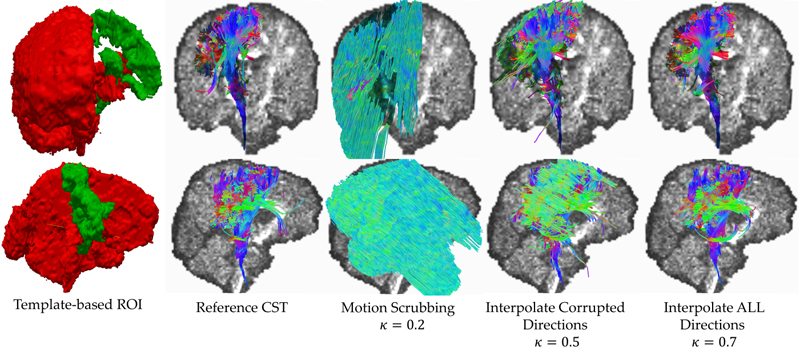
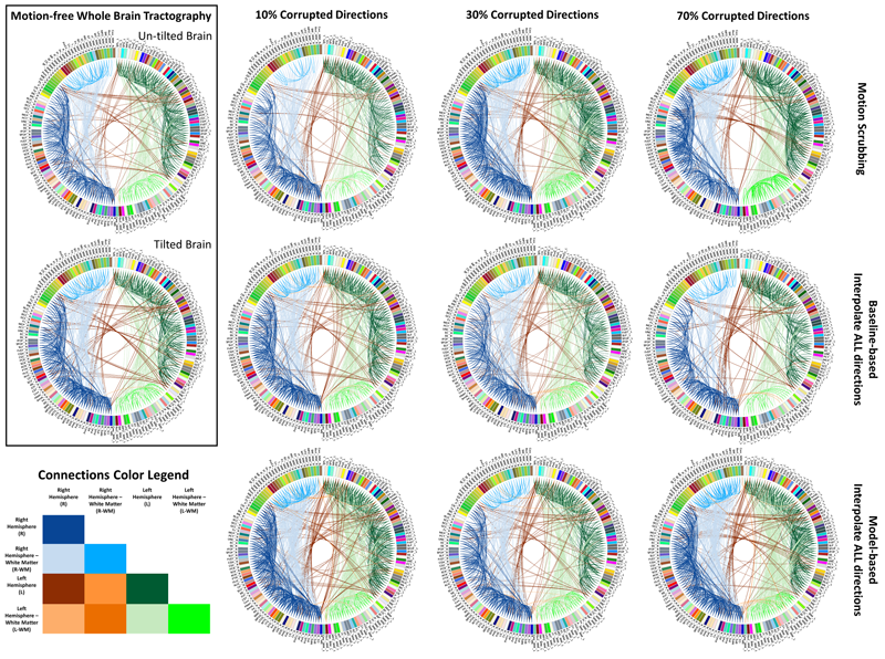
Unlike anatomical MRI where subject motion can most often be assessed by quick visual quality control, the detection, characterization and evaluation of the impact of motion in diffusion imaging are challenging issues due to the sensitivity of diffusion weighted imaging (DWI) to motion originating from vibration, cardiac pulsation, breathing and head movement. Post-acquisition motion correction is widely performed, e.g. using the open-source DTIprep software [1,2] or TORTOISE [3], but in particular in high angular resolution diffusion imaging (HARDI), users often do not fully understand the consequences of different types of correction schemes on the final analysis, and whether those choices may introduce confounding factors when comparing populations. Although there is excellent theoretical work on the number of directional DWI and its effect on the quality and crossing fiber resolution of orientation distribution functions (ODF), standard users lack clear guidelines and recommendations in practical settings. This research investigates motion correction using transformation and interpolation of affected DWI directions versus the exclusion of subsets of DWI’s, and its effects on diffusion measurements on the reconstructed fiber orientation diffusion functions and on the estimated fiber orientations. The various effects are systematically studied via a newly developed synthetic phantom and also on real HARDI data.
![]()
S. Elhabian, Y. Gur, J. Piven, M. Styner, I. Leppert, G. Bruce Pike, G. Gerig.
“Motion is inevitable: The impact of motion correction schemes on hardi reconstructions,” In Proceedings of the MICCAI 2014 Workshop on Computational Diffusion MRI, September, 2014.
![]()
S.Y. Elhabian, Y. Gur, C. Vachet, J. Piven, M.A. Styner, I.R. Leppert, B. Pike, G. Gerig.
“Subject-Motion Correction in HARDI Acquisitions: Choices and Consequences,” In Frontiers in Neurology - Brain Imaging Methods, 2014.
DOI: 10.3389/fneur.2014.00240
Page 1 of 9