
SCI Publications
2024
E. Kwan, E. ghafoori, W. Good, M. Regouski, B. Moon, J. Fish, E. Hsu, I. Polejaeva, R.S. Macleod, D. Dosdall, R. Ranjan.
“Diffuse Functional and Structural Abnormalities in Fibrosis: Potential Structural Basis for Sustaining Atrial Fibrillation,” In Circulation, Vol. 150, pp. A4136863--A4136863. 2024.
2023
![]()
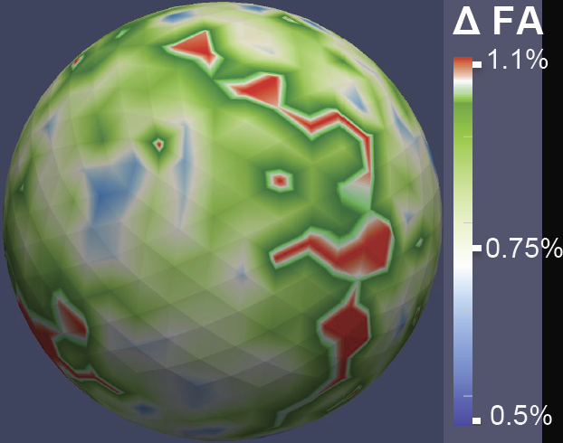
R. Kamali, E. Kwan, M. Regouski, T.J. Bunch, D.J. Dosdall, E. Hsu, R. S. Macleod, I. Polejaeva, R. Ranjan.
“Contribution of atrial myofiber architecture to atrial fibrillation,” In PLOS ONE, Vol. 18, No. 1, Public Library of Science, pp. 1--16. Jan, 2023.
DOI: 10.1371/journal.pone.0279974
Background
The role of fiber orientation on a global chamber level in sustaining atrial fibrillation (AF) is unknown. The goal of this study was to correlate the fiber direction derived from Diffusion Tensor Imaging (DTI) with AF inducibility.
Transgenic goats with cardiac-specific overexpression of constitutively active TGF-β1 (n = 14) underwent AF inducibility testing by rapid pacing in the left atrium. We chose a minimum of 10 minutes of sustained AF as a cut-off for AF inducibility. Explanted hearts underwent DTI to determine the fiber direction. Using tractography data, we clustered, visualized, and quantified the fiber helix angles in 8 different regions of the left atrial wall using two reference vectors defined based on anatomical landmarks.
Sustained AF was induced in 7 out of 14 goats. The mean helix fiber angles in 7 out of 8 selected regions were statistically different (P-Value < 0.05) in the AF inducible group. The average fractional anisotropy (FA) and the mean diffusivity (MD) were similar in the two groups with FA of 0.32±0.08 and MD of 8.54±1.72 mm2/s in the non-inducible group and FA of 0.31±0.05 (P-value = 0.90) and MD of 8.68±1.60 mm2/s (P-value = 0.88) in the inducible group.
DTI based fiber direction shows significant variability across subjects with a significant difference between animals that are AF inducible versus animals that are not inducible. Fiber direction might be contributing to the initiation and sustaining of AF, and its role needs to be investigated further.
2022
![]()
L. Zhou, M. Fan, C. Hansen, C. R. Johnson, D. Weiskopf.
“A Review of Three-Dimensional Medical Image Visualization,” In Health Data Science, Vol. 2022, 2022.
DOI: https://doi.org/10.34133/2022/9840519
Importance. Medical images are essential for modern medicine and an important research subject in visualization. However, medical experts are often not aware of the many advanced three-dimensional (3D) medical image visualization techniques that could increase their capabilities in data analysis and assist the decision-making process for specific medical problems. Our paper provides a review of 3D visualization techniques for medical images, intending to bridge the gap between medical experts and visualization researchers. Highlights. Fundamental visualization techniques are revisited for various medical imaging modalities, from computational tomography to diffusion tensor imaging, featuring techniques that enhance spatial perception, which is critical for medical practices. The state-of-the-art of medical visualization is reviewed based on a procedure-oriented classification of medical problems for studies of individuals and populations. This paper summarizes free software tools for different modalities of medical images designed for various purposes, including visualization, analysis, and segmentation, and it provides respective Internet links. Conclusions. Visualization techniques are a useful tool for medical experts to tackle specific medical problems in their daily work. Our review provides a quick reference to such techniques given the medical problem and modalities of associated medical images. We summarize fundamental techniques and readily available visualization tools to help medical experts to better understand and utilize medical imaging data. This paper could contribute to the joint effort of the medical and visualization communities to advance precision medicine.
2019
![]()
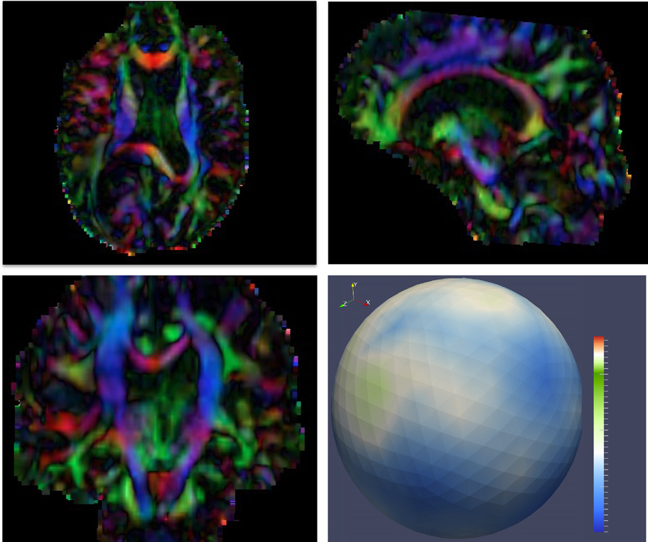
M. Han, I. Wald, W. Usher, Q. Wu, F. Wang, V. Pascicci, C. D. Hansen, C. R. Johnson.
“Ray Tracing Generalized Tube Primitives: Method and Applications,” In Computer Graphics Forum, Vol. 38, No. 3, John Wiley & Sons Ltd., 2019.
We present a general high-performance technique for ray tracing generalized tube primitives. Our technique efficiently supports tube primitives with fixed and varying radii, general acyclic graph structures with bifurcations, and correct transparency with interior surface removal. Such tube primitives are widely used in scientific visualization to represent diffusion tensor imaging tractographies, neuron morphologies, and scalar or vector fields of 3D flow. We implement our approach within the OSPRay ray tracing framework, and evaluate it on a range of interactive visualization use cases of fixed- and varying-radius streamlines, pathlines, complex neuron morphologies, and brain tractographies. Our proposed approach provides interactive, high-quality rendering, with low memory overhead.
2018
O. Abdullah, L. Dai, J. Tippetts, B. Zimmerman, A. Van Hoek, S. Joshi, E. Hsu.
“High resolution and high field diffusion MRI in the visual system of primates (P3.086),” In Neurology, Vol. 90, No. 15 Supplement, Wolters Kluwer Health, Inc, 2018.
ISSN: 0028-3878
Objective: Establishing a primate multiscale genetic brain network linking key microstructural brain components to social behavior remains an elusive goal.
Background: Diffusion MRI, which quantifies the magnitude and anisotropy of water diffusion in brain tissues, offers unparalleled opportunity to link the macroconnectome (resolution of ~0.5mm) to histological-based microconnectome at synaptic resolution.
Design/Methods: We tested the hypothesis that the simplest (and most clinically-used) reconstruction technique (known as diffusion tensor imaging, DTI) will yield similar brain connectivity patterns in the visual system (from optic chiasm to visual cortex) compared to more sophisticated and accurate reconstruction methods including diffusion spectrum imaging (DSI), q-ball imaging (QBI), and generalized q-sampling imaging. We obtained high resolution diffusion MRI data on ex vivo brain from Macaca fascicularis: MRI 7T, resolution 0.5 mm isotropic, 515 diffusion volumes up to b-value (aka diffusion sensitivity) of 40,000 s/mm2 with scan time ~100 hrs.
Results: Tractography results show that despite the limited ability of DTI to resolve crossing fibers at the optic chiasm, DTI-based tracts mapped to the known projections of layers in lateral geniculate nucleus and to the primary visual cortex. The other reconstructions were superior in localized regions for resolving crossing regions.
Conclusions: In conclusion, despite its simplifying assumptions, DTI-based fiber tractography can be used to generate accurate brain connectivity maps that conform to established neuroanatomical features in the visual system.
![]()
D. N. Anderson, B. Osting, J. Vorwerk, A. D Dorval, C. R Butson.
“Optimized programming algorithm for cylindrical and directional deep brain stimulation electrodes,” In Journal of Neural Engineering, Vol. 15, No. 2, pp. 026005. 2018.
Objective. Deep brain stimulation (DBS) is a growing treatment option for movement and psychiatric disorders. As DBS technology moves toward directional leads with increased numbers of smaller electrode contacts, trial-and-error methods of manual DBS programming are becoming too time-consuming for clinical feasibility. We propose an algorithm to automate DBS programming in near real-time for a wide range of DBS lead designs. Approach. Magnetic resonance imaging and diffusion tensor imaging are used to build finite element models that include anisotropic conductivity. The algorithm maximizes activation of target tissue and utilizes the Hessian matrix of the electric potential to approximate activation of neurons in all directions. We demonstrate our algorithm's ability in an example programming case that targets the subthalamic nucleus (STN) for the treatment of Parkinson's disease for three lead designs: the Medtronic 3389 (four cylindrical contacts), the direct STNAcute (two cylindrical contacts, six directional contacts), and the Medtronic-Sapiens lead (40 directional contacts). Main results. The optimization algorithm returns patient-specific contact configurations in near real-time—less than 10 s for even the most complex leads. When the lead was placed centrally in the target STN, the directional leads were able to activate over 50% of the region, whereas the Medtronic 3389 could activate only 40%. When the lead was placed 2 mm lateral to the target, the directional leads performed as well as they did in the central position, but the Medtronic 3389 activated only 2.9% of the STN. Significance. This DBS programming algorithm can be applied to cylindrical electrodes as well as novel directional leads that are too complex with modern technology to be manually programmed. This algorithm may reduce clinical programming time and encourage the use of directional leads, since they activate a larger volume of the target area than cylindrical electrodes in central and off-target lead placements.
2017
![]()
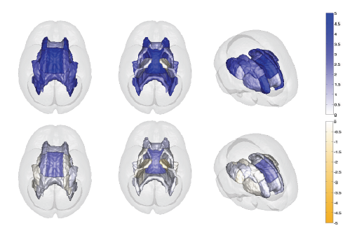
J. J. Wolff, M. R. Swanson, J. T. Elison, G. Gerig, J. R. Pruett, M. A. Styner, C. Vachet, K. N. Botteron, S. R. Dager, A. M. Estes, H. C. Hazlett, R. T. Schultz, M. D. Shen, L. Zwaigenbaum, J. Piven.
“Neural circuitry at age 6~months associated with later repetitive behavior and sensory responsiveness in autism,” In Molecular Autism, Vol. 8, No. 1, Springer Nature, March, 2017.
DOI: 10.1186/s13229-017-0126-z
Background
Restricted and repetitive behaviors are defining features of autism spectrum disorder (ASD). Under revised diagnostic criteria for ASD, this behavioral domain now includes atypical responses to sensory stimuli. To date, little is known about the neural circuitry underlying these features of ASD early in life.
Methods
Longitudinal diffusion tensor imaging data were collected from 217 infants at high familial risk for ASD. Forty-four of these infants were diagnosed with ASD at age 2. Targeted cortical, cerebellar, and striatal white matter pathways were defined and measured at ages 6, 12, and 24 months. Dependent variables included the Repetitive Behavior Scale-Revised and the Sensory Experiences Questionnaire.
Results
Among children diagnosed with ASD, repetitive behaviors and sensory response patterns were strongly correlated, even when accounting for developmental level or social impairment. Longitudinal analyses indicated that the genu and cerebellar pathways were significantly associated with both repetitive behaviors and sensory responsiveness but not social deficits. At age 6 months, fractional anisotropy in the genu significantly predicted repetitive behaviors and sensory responsiveness at age 2. Cerebellar pathways significantly predicted later sensory responsiveness. Exploratory analyses suggested a possible disordinal interaction based on diagnostic status for the association between fractional anisotropy and repetitive behavior.
Conclusions
Our findings suggest that restricted and repetitive behaviors contributing to a diagnosis of ASD at age 2 years are associated with structural properties of callosal and cerebellar white matter pathways measured during infancy and toddlerhood. We further identified that repetitive behaviors and unusual sensory response patterns co-occur and share common brain-behavior relationships. These results were strikingly specific given the absence of association between targeted pathways and social deficits.
2015
![]()
S. Pujol, W. Wells, C. Pierpaoli, C. Brun, J. Gee, G. Cheng, B. Vemuri, O. Commowick, S. Prima, A. Stamm, M. Goubran, A. Khan, T. Peters, P. Neher, K. H. Maier-Hein, Y. Shi, A. Tristan-Vega, G. Veni, R. Whitaker, M. Styner, C.F. Westin, S. Gouttard, I. Norton, L. Chauvin, H. Mamata, G. Gerig, A. Nabavi, A. Golby,, R. Kikinis.
“The DTI Challenge: Toward Standardized Evaluation of Diffusion Tensor Imaging Tractography for Neurosurgery,” In Journal of Neuroimaging, Wiley, August, 2015.
DOI: 10.1111/jon.12283

Diffusion tensor imaging (DTI) tractography reconstruction of white matter pathways can help guide brain tumor resection. However, DTI tracts are complex mathematical objects and the validity of tractography-derived information in clinical settings has yet to be fully established. To address this issue, we initiated the DTI Challenge, an international working group of clinicians and scientists whose goal was to provide standardized evaluation of tractography methods for neurosurgery. The purpose of this empirical study was to evaluate different tractography techniques in the first DTI Challenge workshop.
METHODS
Eight international teams from leading institutions reconstructed the pyramidal tract in four neurosurgical cases presenting with a glioma near the motor cortex. Tractography methods included deterministic, probabilistic, filtered, and global approaches. Standardized evaluation of the tracts consisted in the qualitative review of the pyramidal pathways by a panel of neurosurgeons and DTI experts and the quantitative evaluation of the degree of agreement among methods.
RESULTS
The evaluation of tractography reconstructions showed a great interalgorithm variability. Although most methods found projections of the pyramidal tract from the medial portion of the motor strip, only a few algorithms could trace the lateral projections from the hand, face, and tongue area. In addition, the structure of disagreement among methods was similar across hemispheres despite the anatomical distortions caused by pathological tissues.
CONCLUSIONS
The DTI Challenge provides a benchmark for the standardized evaluation of tractography methods on neurosurgical data. This study suggests that there are still limitations to the clinical use of tractography for neurosurgical decision making.
![]()
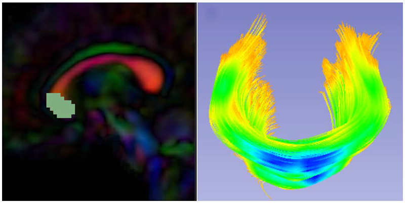
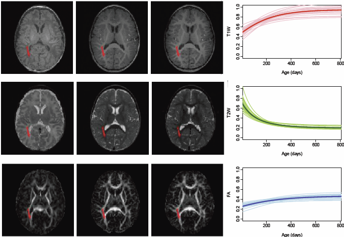
N. Sadeghi, J. H. Gilmore , G. Gerig.
“Modeling Brain Growth and Development,” In Brain, Vol. 1, pp. 429-436. 2015.
DOI: 10.1016/B978-0-12-397025-1.00314-6
![]()
J. J. Wolff, G. Gerig, J. D. Lewis, T. Soda, M. A. Styner, C. Vachet, K. N. Botteron, J. T. Elison, S. R. Dager, A. M. Estes, H. C. Hazlett, R. T. Schultz, L. Zwaigenbaum, J. Piven.
“Altered corpus callosum morphology associated with autism over the first 2 years of life,” In Brain, 2015.
DOI: 10.1093/brain/awv118

2014
![]()
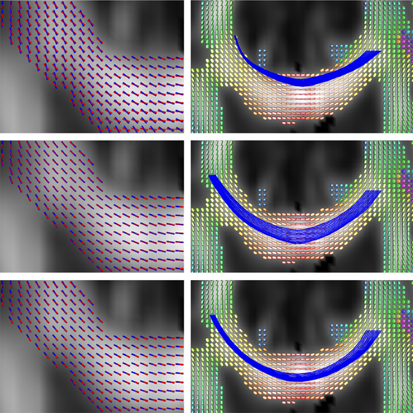
G. Adluru, Y. Gur, J. Anderson, L. Richards, N. Adluru, E. DiBella.
“Assessment of white matter microstructure in stroke patients using NODDI,” In Proceedings of the 2014 IEEE Int. Conf. Engineering and Biology Society (EMBC), 2014.
Diffusion weighted imaging (DWI) is widely used to study changes in white matter following stroke. In various studies employing diffusion tensor imaging (DTI) and high angular resolution diffusion imaging (HARDI) modalities, it has been shown that fractional anisotropy (FA), mean diffusivity (MD), and generalized FA (GFA) can be used as measures of white matter tract integrity in stroke patients. However, these measures may be non-specific, as they do not directly delineate changes in tissue microstructure. Multi-compartment models overcome this limitation by modeling DWI data using a set of indices that are directly related to white matter microstructure. One of these models which is gaining popularity, is neurite orientation dispersion and density imaging (NODDI). This model uses conventional single or multi-shell HARDI data to describe fiber orientation dispersion as well as densities of different tissue types in the imaging voxel. In this paper, we apply for the first time the NODDI model to 4-shell HARDI stroke data. By computing NODDI indices over the entire brain in two stroke patients, and comparing tissue regions in ipsilesional and contralesional hemispheres, we demonstrate that NODDI modeling provides specific information on tissue microstructural changes. We also introduce an information theoretic analysis framework to investigate the non-local effects of stroke in the white matter. Our initial results suggest that the NODDI indices might be more specific markers of white matter reorganization following stroke than other measures previously used in studies of stroke recovery.
![]()
X. Hao, K. Zygmunt, R.T. Whitaker, P.T. Fletcher.
“Improved Segmentation of White Matter Tracts with Adaptive Riemannian Metrics,” In Medical Image Analysis, Vol. 18, No. 1, pp. 161--175. Jan, 2014.
DOI: 10.1016/j.media.2013.10.007
PubMed ID: 24211814
Keywords: Conformal factor, Diffusion tensor imaging, Front-propagation, Geodesic, Riemannian manifold
![]()
I. Oguz, M. Farzinfar, J. Matsui, F. Budin, Z. Liu, G. Gerig, H.J. Johnson, M.A. Styner.
“DTIPrep: Quality Control of Diffusion-Weighted Images,” In Frontiers in Neuroinformatics, Vol. 8, No. 4, 2014.
DOI: 10.3389/fninf.2014.00004
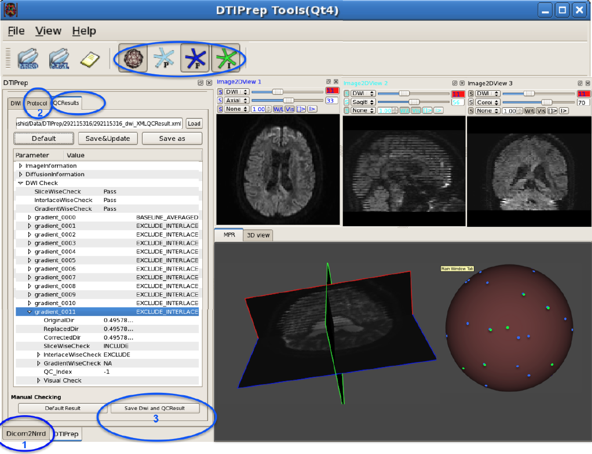
In the last decade, diffusion MRI (dMRI) studies of the human and animal brain have been used to investigate a multitude of pathologies and drug-related effects in neuroscience research. Study after study identifies white matter (WM) degeneration as a crucial biomarker for all these diseases. The tool of choice for studying WM is dMRI. However, dMRI has inherently low signal-to-noise ratio and its acquisition requires a relatively long scan time; in fact, the high loads required occasionally stress scanner hardware past the point of physical failure. As a result, many types of artifacts implicate the quality of diffusion imagery. Using these complex scans containing artifacts without quality control (QC) can result in considerable error and bias in the subsequent analysis, negatively affecting the results of research studies using them. However, dMRI QC remains an under-recognized issue in the dMRI community as there are no user-friendly tools commonly available to comprehensively address the issue of dMRI QC. As a result, current dMRI studies often perform a poor job at dMRI QC.
Thorough QC of diffusion MRI will reduce measurement noise and improve reproducibility, and sensitivity in neuroimaging studies; this will allow researchers to more fully exploit the power of the dMRI technique and will ultimately advance neuroscience. Therefore, in this manuscript, we present our open-source software, DTIPrep, as a unified, user friendly platform for thorough quality control of dMRI data. These include artifacts caused by eddy-currents, head motion, bed vibration and pulsation, venetian blind artifacts, as well as slice-wise and gradient-wise intensity inconsistencies. This paper summarizes a basic set of features of DTIPrep described earlier and focuses on newly added capabilities related to directional artifacts and bias analysis.
Keywords: diffusion MRI, Diffusion Tensor Imaging, Quality control, Software, open-source, preprocessing
![]()
N. Sadeghi, J.H. Gilmore, W. Lin, G. Gerig.
“Normative Modeling of Early Brain Maturation from Longitudinal DTI Reveals Twin-Singleton Differences,” In Proceeding of the 2014 Joint Annual Meeting ISMRM-ESMRMB, pp. (accepted). 2014.
Early brain development of white matter is characterized by rapid organization and structuring. Magnetic Resonance diffusion tensor imaging (MR-DTI) provides the possibility of capturing these changes non-invasively by following individuals longitudinally in order to better understand departures from normal brain development in subjects at risk for mental illness [1]. Longitudinal imaging of individuals suggests the use of 4D (3D, time) image analysis and longitudinal statistical modeling [3].
![]()
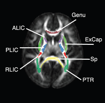
A.R. Verde, F. Budin, J.-B. Berger, A. Gupta, M. Farzinfar, A. Kaiser, M. Ahn, H. Johnson, J. Matsui, H.C. Hazlett, A. Sharma, C. Goodlett, Y. Shi, S. Gouttard, C. Vachet, J. Piven, H. Zhu, G. Gerig, M. Styner.
“UNC-Utah NA-MIC framework for DTI fiber tract analysis,” In Frontiers in Neuroinformatics, Vol. 7, No. 51, January, 2014.
DOI: 10.3389/fninf.2013.00051
Keywords: neonatal neuroimaging, white matter pathways, magnetic resonance imaging, diffusion tensor imaging, diffusion imaging quality control, DTI atlas building
2013
![]()
A. Daducci, E.J. Canales-Rodriguez, M. Descoteaux, E. Garyfallidis, Y. Gur, Y.-C Lin, M. Mani, S. Merlet, M. Paquette, A. Ramirez-Manzanares, M. Reisert, P.R. Rodrigues, F. Sepehrband, E. Caruyer, J. Choupan, R. Deriche, M. Jacob, G. Menegaz, V. Prckovska, M. Rivera, Y. Wiaux, J.-P. Thiran.
“Quantitative comparison of reconstruction methods for intra-voxel fiber recovery from diffusion MRI,” In IEEE Transactions on Medical Imaging, Vol. 33, No. 2, pp. 384--399. 2013.
ISSN: 0278-0062
DOI: 10.1109/TMI.2013.2285500
Validation is arguably the bottleneck in the diffusion MRI community. This paper evaluates and compares 20 algorithms for recovering the local intra-voxel fiber structure from diffusion MRI data and is based on the results of the "HARDI reconstruction challenge" organized in the context of the "ISBI 2012" conference. Evaluated methods encompass a mixture of classical techniques well-known in the literature such as Diffusion Tensor, Q-Ball and Diffusion Spectrum imaging, algorithms inspired by the recent theory of compressed sensing and also brand new approaches proposed for the first time at this contest. To quantitatively compare the methods under controlled conditions, two datasets with known ground-truth were synthetically generated and two main criteria were used to evaluate the quality of the reconstructions in every voxel: correct assessment of the number of fiber populations and angular accuracy in their orientation. This comparative study investigates the behavior of every algorithm with varying experimental conditions and highlights strengths and weaknesses of each approach.
![]()
M. Farzinfar, Y. Li, A.R. Verde, I. Oguz, G. Gerig, M.A. Styner.
“DTI Quality Control Assessment via Error Estimation From Monte Carlo Simulations,” In Proceedings of SPIE 8669, Medical Imaging 2013: Image Processing, Vol. 8669, 2013.
DOI: 10.1117/12.2006925
PubMed ID: 23833547
PubMed Central ID: PMC3702180
![]()
M. Farzinfar, I. Oguz, R.G. Smith, A.R. Verde, C. Dietrich, A. Gupta, M.L. Escolar, J. Piven, S. Pujol, C. Vachet, S. Gouttard, G. Gerig, S. Dager, R.C. McKinstry, S. Paterson, A.C. Evans, M.A. Styner.
“Diffusion imaging quality control via entropy of principal direction distribution,” In NeuroImage, Vol. 82, pp. 1--12. 2013.
ISSN: 1053-8119
DOI: 10.1016/j.neuroimage.2013.05.022
Keywords: Diffusion magnetic resonance imaging, Diffusion tensor imaging, Quality assessment, Entropy
![]()
N. Sadeghi, M.W. Prastawa, P.T. Fletcher, C. Vachet, Bo Wang, J.H. Gilmore, G. Gerig.
“Multivariate Modeling of Longitudinal MRI in Early Brain Development with Confidence Measures,” In Proceedings of the 2013 IEEE 10th International Symposium on Biomedical Imaging (ISBI), pp. 1400--1403. 2013.
DOI: 10.1109/ISBI.2013.6556795
![]()
N. Sadeghi, M.W. Prastawa, P.T. Fletcher, J. Wolff, J.H. Gilmore, G. Gerig.
“Regional characterization of longitudinal DT-MRI to study white matter maturation of the early developing brain,” In NeuroImage, Vol. 68, pp. 236--247. March, 2013.
DOI: 10.1016/j.neuroimage.2012.11.040
PubMed ID: 23235270
Page 1 of 4