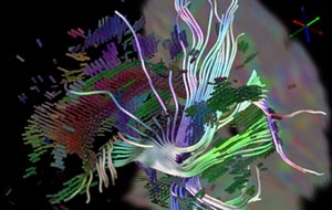
Diffusion tensor MRI visualization is a growing field of research. The scanners are collecting better data all the time, and doctors and scientists are constantly discovering new applications for this data. However, unlike scalar and vector data, high-dimensional tensors are not always intuitive to visualize. When devising new strategies for DT-MRI visualization, it is important to understand both what exactly it is that is being measured and what insights the doctors and scientists are hoping to gain from the data.
The success of diffusion magnetic resonance imaging (MRI) is deeply rooted in the powerful concept that during their random, diffusion-driven displacements, molecules probe tissue structure at a microscopic scale well beyond the usual image resolution. As diffusion is truly a three dimensional process, molecular mobility in tissues may be anisotropic, as in brain white matter. With diffusion tensor imaging (DTI), diffusion anisotropy effects can be fully extracted, characterized, and exploited, providing even more exquisite details on tissue microstructure. The most advanced application is certainly that of fiber tracking in the brain, which, in combination with functional MRI, might open a window on the important issue of connectivity. DTI has also been used to demonstrate subtle abnormalities in a variety of diseases (including stroke, multiple sclerosis, dyslexia, and schizophrenia) and is currently becoming part of many routine clinical protocols.
 Diffusion tensor MRI visualization is a growing field of research. The scanners are collecting better data all the time, and doctors and scientists are constantly discovering new applications for this data. However, unlike scalar and vector data, high-dimensional tensors are not always intuitive to visualize. When devising new strategies for DT-MRI visualization, it is important to understand both what exactly it is that is being measured and what insights the doctors and scientists are hoping to gain from the data.