Region Segmentation using HMT
The first step for EM image segmentation is to automatically detecting cell membranes using supervised machine learning, which results in probability maps of cell boundaries. Then region segmentation is generated that assigns each neuron cell a unique integer label. In this section, we focus on this region segmentation step. One simple solution is thresholding the boundary probability maps. However, since the boundary predictions are usually not perfect, it is usually difficult or even impossible to find a good thresholding level.
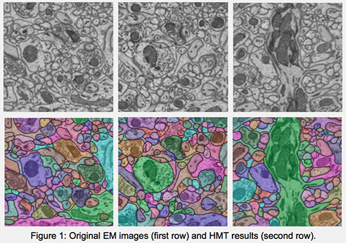
We propose a hierarchical approach to the region segmentation problem, namely HMT [1,2]. First, we generate initial superpixel segmentation of an image using the watershed algorithm. Next, we use a merge tree structure to represent the merging order of the superpixels. Then we train a boundary classifier to predict how likely a pair of regions should merge, based on which we infer the final segmentation from the tree under certain consistency constraint. Our approach was the stateoftheart in the ISBI 2012 and 2013 EM segmentation challenge. Figure 1 shows some sample segmentation results.
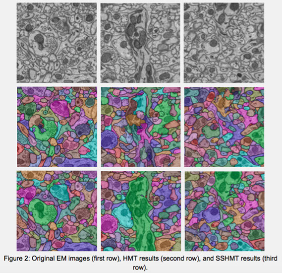
Supervised learning of the boundary classifier demands considerable ground truth data, which are costly to collect from EM images. Human experts are expected to manually label every pixel in an image. We propose a semisupervised approach, namely SSHMT [2], to reduce this demand. Based on the merge tree structure, we develop a differentiable unsupervised loss term that enforces consistent predictions from the learned classifier. Then we propose a Bayesian model that combines the supervised and the unsupervised information for probabilistic learning. The experimental results show that by using a subset of only a small portion of the entire ground truth data, our semisupervised approach consistently performs close to the fully supervised method trained with the full labeled data set. Figure 2 shows some sample segmentation results comparison.
References:
[1] Watershed merge tree classification for electron microscopy image segmentation. ICPR 2012.
[2] A modular approach to 3D electron microscopy image segmentation. Journal of Neuroscience Methods, 2014.
[3] SSHMT: Semisupervised hierarchical merge tree for electron microscopy image segmentation. ECCV 2016.
© 2014
Scientific Computing and Imaging Institute