SCIENTIFIC COMPUTING AND IMAGING INSTITUTEat the University of Utah
An internationally recognized leader in visualization, scientific computing, and image analysis
SCI Publications
2021
Integrin-Based Mechanosensing through Conformational Deformation
T.P. Driscoll, T.C. Bidone, S.J. Ahn, A. Yu, A. Groisman, G.A. Voth, M.A. Schwartz.
“Integrin-Based Mechanosensing through Conformational Deformation,” In Biophysical Journal, 2021.
DOI: https://doi.org/10.1016/j.bpj.2021.09.010
ABSTRACT
Conversion of integrins from low to high affinity states, termed activation, is important in biological processes including immunity, hemostasis, angiogenesis and embryonic development. Integrin activation is regulated by large-scale conformational transitions from closed, low affinity states to open, high affinity states. While it has been suggested that substrate stiffness shifts the conformational equilibrium of integrin and governs its unbinding, here we address the role of integrin conformational activation in cellular mechanosensing. Comparison of WT vs activating mutants of integrin αVβ3 show that activating mutants shift cell spreading, FAK activation, traction stress and force on talin toward high stiffness values at lower stiffness. Although all activated integrin mutants showed equivalent binding affinity for soluble ligands, the β3 S243E mutant showed the strongest shift in mechanical responses. To understand this behavior, we used coarse-grained computational models derived from molecular level information. The models predicted that wild type integrin αVβ3 displaces under force, and that activating mutations shift the required force toward lower values, with S243E showing the strongest effect. Cellular stiffness sensing thus correlates with computed effects of force on integrin conformation. Together, these data identify a role for force-induced integrin conformational deformation in cellular mechanosensing.
2014
Extracellular matrix density regulates the rate of neovessel growth and branching in sprouting angiogenesis
L.T. Edgar, C.J. Underwood, J.E. Guilkey, J.B. Hoying, J.A. Weiss.
“Extracellular matrix density regulates the rate of neovessel growth and branching in sprouting angiogenesis,” In PLOS one, Vol. 9, No. 1, 2014.
DOI: 10.1371/journal.pone.0085178
ABSTRACT
×
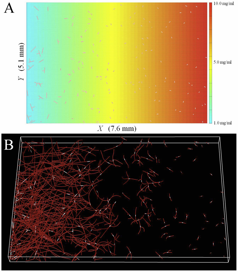
Angiogenesis is regulated by the local microenvironment, including the mechanical interactions between neovessel sprouts and the extracellular matrix (ECM). However, the mechanisms controlling the relationship of mechanical and biophysical properties of the ECM to neovessel growth during sprouting angiogenesis are just beginning to be understood. In this research, we characterized the relationship between matrix density and microvascular topology in an in vitro 3D organ culture model of sprouting angiogenesis. We used these results to design and calibrate a computational growth model to demonstrate how changes in individual neovessel behavior produce the changes in vascular topology that were observed experimentally. Vascularized gels with higher collagen densities produced neovasculatures with shorter vessel lengths, less branch points, and reduced network interconnectivity. The computational model was able to predict these experimental results by scaling the rates of neovessel growth and branching according to local matrix density. As a final demonstration of utility of the modeling framework, we used our growth model to predict several scenarios of practical interest that could not be investigated experimentally using the organ culture model. Increasing the density of the ECM significantly reduced angiogenesis and network formation within a 3D organ culture model of angiogenesis. Increasing the density of the matrix increases the stiffness of the ECM, changing how neovessels are able to deform and remodel their surroundings. The computational framework outlined in this study was capable of predicting this observed experimental behavior by adjusting neovessel growth rate and branching probability according to local ECM density, demonstrating that altering the stiffness of the ECM via increasing matrix density affects neovessel behavior, thereby regulated vascular topology during angiogenesis.
Formation of microvascular networks: role of stromal interactions directing angiogenic growth
J.B. Hoying, U. Utzinger, J.A. Weiss.
“Formation of microvascular networks: role of stromal interactions directing angiogenic growth,” In Microcirculation, Vol. 21, No. 4, pp. 278--289. May, 2014.
DOI: 10.1111/micc.12115
PubMed ID: 24447042
PubMed Central ID: PMC4032604
ABSTRACT
×
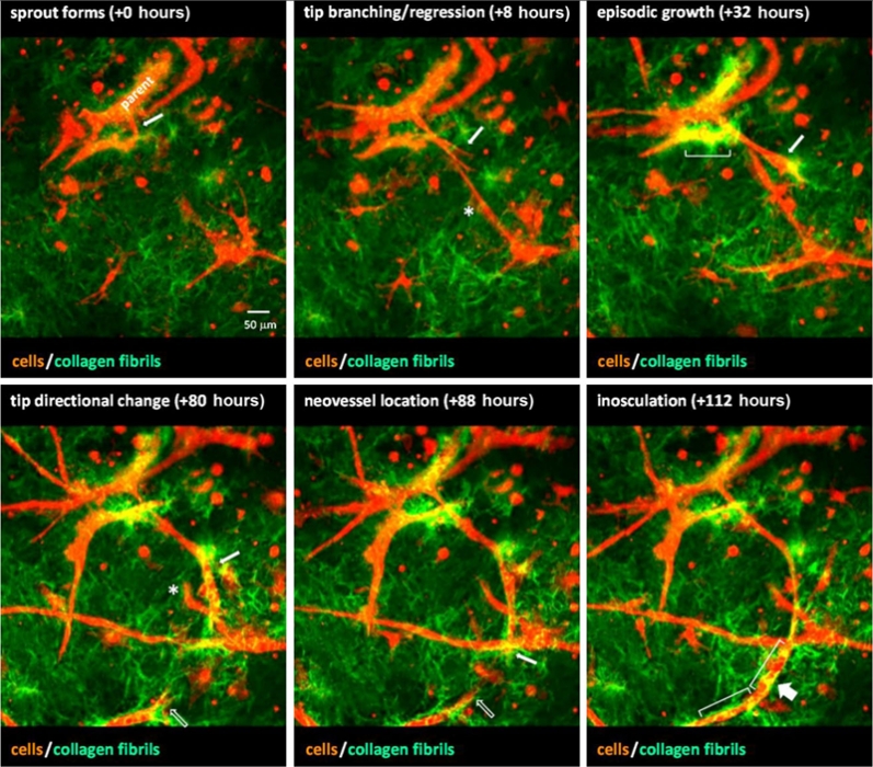
In the adult, angiogenesis leads to an expanded microvascular network as new vessel segments are added to an existing microcirculation. Necessarily, growing neovessels must navigate through tissue stroma as they locate and grow toward other vessel elements. We have a growing body of evidence demonstrating that angiogenic neovessels reciprocally interact with the interstitial matrix of the stroma resulting in directed neovascular growth during angiogenesis. Given the compliance and the viscoelastic properties of collagen, neovessel guidance by the stroma is likely due to compressive strain transverse to the direction of primary tensile forces present during active tissue deformation. Similar stromal strains control the final network topology of the new microcirculation, including the distribution of arterioles, capillaries, and venules. In this case, stromal-derived stimuli must be present during the post-angiogenesis remodeling and maturation phases of neovascularization to have this effect. Interestingly, the preexisting organization of vessels prior to the start of angiogenesis has no lasting influence on the final, new network architecture. Combined, the evidence describes interplay between angiogenic neovessels and stroma that is important in directed neovessel growth and invasion. This dynamic is also likely a mechanism by which global tissue forces influence vascular form and function.
Cell-generated traction forces and the resulting matrix deformation modulate microvascular alignment and growth during angiogenesis
C.J. Underwood, L.T. Edgar, J.B. Hoying, J.A. Weiss.
“Cell-generated traction forces and the resulting matrix deformation modulate microvascular alignment and growth during angiogenesis,” In American Journal of Physiology: Heart and Circulatory Physiology, Vol. 307, No. H152-H164, 2014.
DOI: 10.1152/ajpheart.00995.2013
PubMed ID: 24816262
PubMed Central ID: PMC4101638
ABSTRACT
×
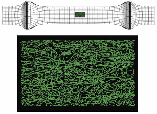
The details of the mechanical factors that modulate angiogenesis remain poorly understood. Previous in vitro studies of angiogenesis using microvessel fragments cultured within collagen constructs demonstrated that neovessel alignment can be induced via mechanical constraint of the boundaries (i.e., boundary conditions). The objective of this study was to investigate the role of mechanical boundary conditions in the regulation of angiogenic alignment and growth in an in vitro model of angiogenesis. Angiogenic microvessels within three-dimensional constructs were subjected to different boundary conditions, thus producing different stress and strain fields during growth. Neovessel outgrowth and orientation were quantified from confocal image data after 6 days. Vascularity and branching decreased as the amount of constraint imposed on the culture increased. In long-axis constrained hexahedral constructs, microvessels aligned parallel to the constrained axis. In contrast, constructs that were constrained along the short axis had random microvessel orientation. Finite element models were used to simulate the contraction of gels under the various boundary conditions and to predict the local strain field experienced by microvessels. Results from the experiments and simulations demonstrated that microvessels aligned perpendicular to directions of compressive strain. Alignment was due to anisotropic deformation of the matrix from cell-generated traction forces interacting with the mechanical boundary conditions. These findings demonstrate that boundary conditions and thus the effective stiffness of the matrix regulate angiogenesis. This study offers a potential explanation for the oriented vascular beds that occur in native tissues and provides the basis for improved control of tissue vascularization in both native tissues and tissue-engineered constructs.
A computational model of in vitro angiogenesis based on extracellular matrix fiber orientation
L.T. Edgar, S.C. Sibole, C.J. Underwood, J.E. Guilkey, J.A. Weiss.
“A computational model of in vitro angiogenesis based on extracellular matrix fiber orientation,” In Computer Methods in Biomechanical and Biomedical Engineering, Vol. 16, No. 7, pp. 790--801. 2013.
DOI: 10.1080/10255842.2012.662678
ABSTRACT
×
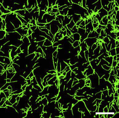
Recent interest in the process of vascularisation within the biomedical community has motivated numerous new research efforts focusing on the process of angiogenesis. Although the role of chemical factors during angiogenesis has been well documented, the role of mechanical factors, such as the interaction between angiogenic vessels and the extracellular matrix, remains poorly understood. In vitro methods for studying angiogenesis exist; however, measurements available using such techniques often suffer from limited spatial and temporal resolutions. For this reason, computational models have been extensively employed to investigate various aspects of angiogenesis. This paper outlines the formulation and validation of a simple and robust computational model developed to accurately simulate angiogenesis based on length, branching and orientation morphometrics collected from vascularised tissue constructs. Microvessels were represented as a series of connected line segments. The morphology of the vessels was determined by a linear combination of the collagen fibre orientation, the vessel density gradient and a random walk component. Excellent agreement was observed between computational and experimental morphometric data over time. Computational predictions of microvessel orientation within an anisotropic matrix correlated well with experimental data. The accuracy of this modelling approach makes it a valuable platform for investigating the role of mechanical interactions during angiogenesis.
2012
Determinants of microvascular network topology in implanted neovasculatures
C.C. Chang, L. Krishnan, S.S. Nunes, K.H. Church, L.T. Edgar, E.D. Boland, J.A. Weiss, S.K. Williams, J.B. Hoying.
“Determinants of microvascular network topology in implanted neovasculatures,” In Arteriosclerosis, Thrombosis, and Vascular Biology, Vol. 32, No. 1, pp. 5--14. 2012.
DOI: 10.1161/ATVBAHA.111.238725
ABSTRACT
×
Objective During neovascularization, the end result is a new functional microcirculation composed of a network of mature microvessels with specific topologies. Although much is known concerning the mechanisms underlying the initiation of angiogenesis, it remains unclear how the final architecture of microcirculatory beds is regulated. To begin to address this, we determined the impact of angiogenic neovessel prepatterning on the final microvascular network topology using a model of implant neovascularization.
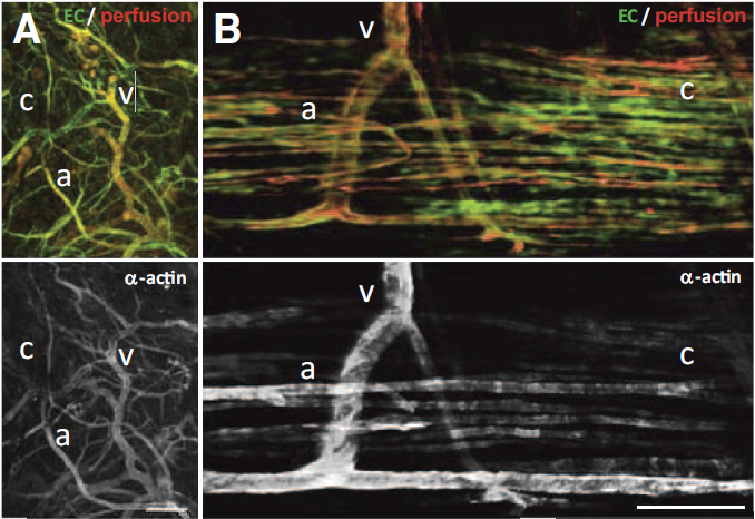
Methods and Results We used 3D direct-write bioprinting or physical constraints in a manner permitting postangiogenesis vascular remodeling and adaptation to pattern angiogenic microvascular precursors (neovessels formed from isolated microvessel segments) in 3D collagen gels before implantation and subsequent network formation. Neovasculatures prepatterned into parallel arrays formed functional networks after 4 weeks postimplantation but lost the prepatterned architecture. However, maintenance of uniaxial physical constraints during postangiogenesis remodeling of the implanted neovasculatures produced networks with aligned microvessels, as well as an altered proportional distribution of arterioles, capillaries, and venules.
Conclusion Here we show that network topology resulting from implanted microvessel precursors is independent from prepatterning of precursors but can be influenced by a patterning stimulus involving tissue deformation during postangiogenesis remodeling and maturation.
2008
Effect of Mechanical Boundary Conditions on Orientation of Angiogenic Microvessels
L. Krishnan, C.J. Underwood, S.A. Maas, B.J. Ellis, T.C. Kode, J.B. Hoying, J.A. Weiss.
“Effect of Mechanical Boundary Conditions on Orientation of Angiogenic Microvessels,” In Cardiovascular Research, Vol. 78, No. 2, pp. 324--332. 2008.
2006
In Vitro Model for Endogenous Optical Signatures of Collagen
N.D. Kirkpatrick, J.B. Hoying, S.K. Botting, J.A. Weiss, U. Utzinger.
“In Vitro Model for Endogenous Optical Signatures of Collagen,” In Journal of Biomedical Optics, Vol. 11, No. 5, September/October, 2006.
Permeability of Human Medial Collateral Ligament Transverse to the Collagen Fiber Direction
J.A. Weiss, B.J. Maakestad.
“Permeability of Human Medial Collateral Ligament Transverse to the Collagen Fiber Direction,” In J. Biomech., Vol. 39, No. 2, pp. 276--283. 2006.
2005
Viscoelastic Properties of the Human Medial Collateral Ligament Under Longitudinal, Transverse and Shear Loading
C. Bonifasi-Lista, S.P. Lake, M. Small, J.A. Weiss.
“Viscoelastic Properties of the Human Medial Collateral Ligament Under Longitudinal, Transverse and Shear Loading,” In J. Orthoped. Res., Vol. 23, No. 1, pp. 67--76. January, 2005.
Computational modeling of multicellular constructs with the Material Point Method
J.E. Guilkey, J.B. Hoying JB, J.A. Weiss.
“Computational modeling of multicellular constructs with the Material Point Method,” In Journal of Biomechanics, pp. (published online). June, 2005.
The Role of Mechanical Stresses in Angiogenesis
Y.T. Shiu, J.A. Weiss, J.B. Hoying, M.N. Iwamoto, I.S. Joung, C.T. Quam.
“The Role of Mechanical Stresses in Angiogenesis,” In CRC Crit. Rev. Biomed. Eng., Vol. 33, No. 5, pp. 431--510. 2005.
2004
Design and Application of a Test System for Viscoelastic Characterization of Collagen Gels
L. Krishnan, J.A. Weiss, M.D. Wessman MD, J.B. Hoying.
“Design and Application of a Test System for Viscoelastic Characterization of Collagen Gels,” In Tiss. Eng., Vol. 10, No. (1-2), pp. 241--252. 2004.