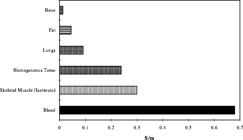
Figure 3: Range of conductivies assigned to different regions of the torso model.
Electric conductivity is the material property of interest in modeling electrocardiographic fields. At a microscopic level, the discrete nature of cell structure dictates that all tissue is anisotropic; however, at a macroscopic level, many tissues can be approximated as having isotropic conductivity in the 0-100 Hz bandwidth that is considered relevant for electrocardiography [17]. The exception is the skeletal muscle, which maintains anisotropic properties even at a macroscopic level. We implemented anisotropy in this model by specifying a 3 x 3 conductivity tensor with different values for each direction of conductivity. Each tetrahedra was automatically assigned a conductivity tensor according to the tissue type to which it belonged. All tissues, except skeletal muscle, were modeled as isotropic and piecewise homogeneous. In the isotropic case, the conductivity tensor reduces to a scalar, while for anisotropic regions it is a 3 x 3 symmetric tensor.

Figure 3: Range of conductivies assigned to different regions of the torso model.
The conductivity values used for the tissue types in this model were taken from the comprehensive review article of Foster and Schwann [16]. Conductivity in living tissue varies sharply with frequency and Figure 3 demonstrates the large range of conductivity values found among the tissues in the torso for the 0-100 Hz range. The homogeneous torso conductivity, .239 S/m, is the mean of the conductivities of the other tissues and was used for the homogeneous torso model.

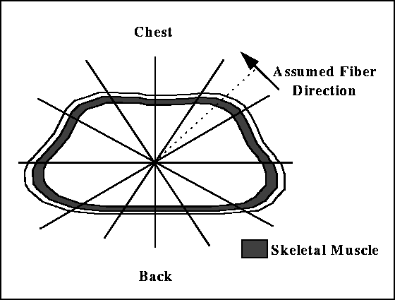
Figure 4: Anisotropic conduction assignment for the skeletal muscle. Fiber direction of the muscle was assumed to be perpendicular to the bisector (shown as dashed line) of each of 12 sectors (shown bounded by solid lines) in the torso cross section.
Within the skeletal muscle that surrounds the thorax, fiber orientation--and hence conductivity--changes constantly. To approximate the resulting directional conductivity, we considered the torso to be a cylinder around which the muscle fibers wrap. We then split the torso into twelve 30 degree sections or slices and assumed the fiber direction of each slice to be perpendicular to the bisector of the slice. Figure 4 shows a schematic of the fiber directions. Anisotropy ratios indicate the relative conductivities along (logitudinal) and perpendicular to (radial) the local fiber direction. Longitudinal to radial conductivity ratios in the simulations ranged from 1:1 (isotropic) through 3:1, 5:1, 7:1 (the value cited in most literature [16]), 10:1 to 15:1.