Invariant Object Descriptors and Matching
Motivation: Whereas object matching and description is an important goal for many medical imaging and computer vision applications, successful object description requires invariance and distinction. The object description methodology must be robust to accommodate various variations in imaging conditions while producing a distinctive characterization of the desired object.
- Pulmonary nodule classification for early diagnosis of lung cancer
- Modeling corpus callosum shape for early detection of autism
Pulmonary nodule classification for early diagnosis of lung cancer
Joint work with: Aly Farag
Lung cancer in the United States account for 30% of all cancer-related deaths, resulting in over 160,000 deaths per year, which is more than the annual deaths for colon, breast, prostate, ovarian and pancreatic cancers combined. The survival of lung cancer is strongly dependent of diagnosis. My colleagues and I at the Computer Vision and Image Processing (CVIP) Lab at the University of Louisville (UofL) developed a CAD system for pulmonary nodule classification based on shape and texture descriptors. We deployed a nonparametric approach for building data-driven models for different types of pulmonary nodules, where shape and texture models have shown promising results.
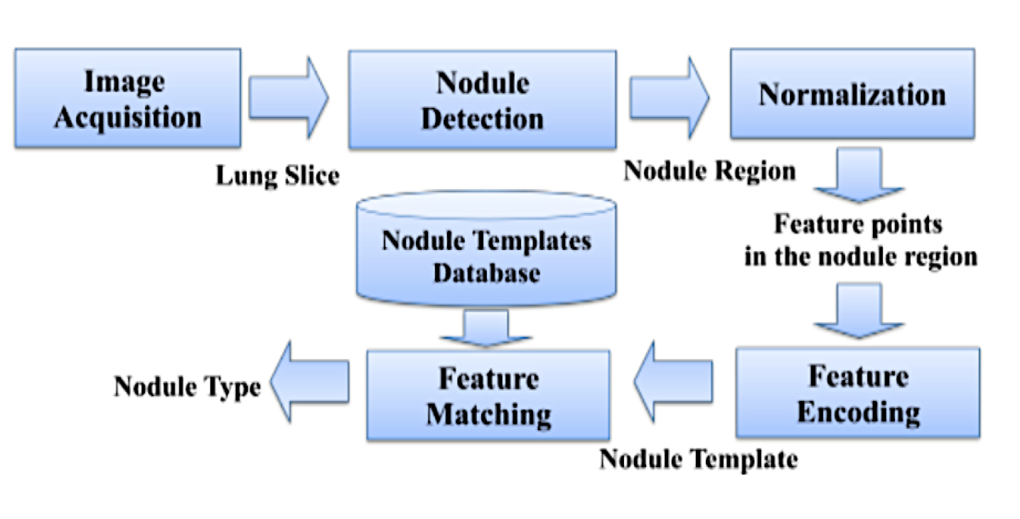
Nodule Classification block-diagram adopted from Daugman’s Iris
Recognition framework.
The Scale Invariant Feature Transform (SIFT) and an adaptation to Daugman’s Iris Recognition algorithm were used for analysis. The SIFT descriptor results were projected to lower-dimensional subspaces using PCA and LDA. Complex Gabor wavelet nodule response obtained from an adapted Daugman Iris Recognition algorithm revealed improvements from the original Daugman binary iris code. This showed that binarized nodule responses (codes) are inadequate for classification since nodules lack texture concentration as seen in the iris, while the SIFT algorithm projected using PCA showed robustness and precision in classification.
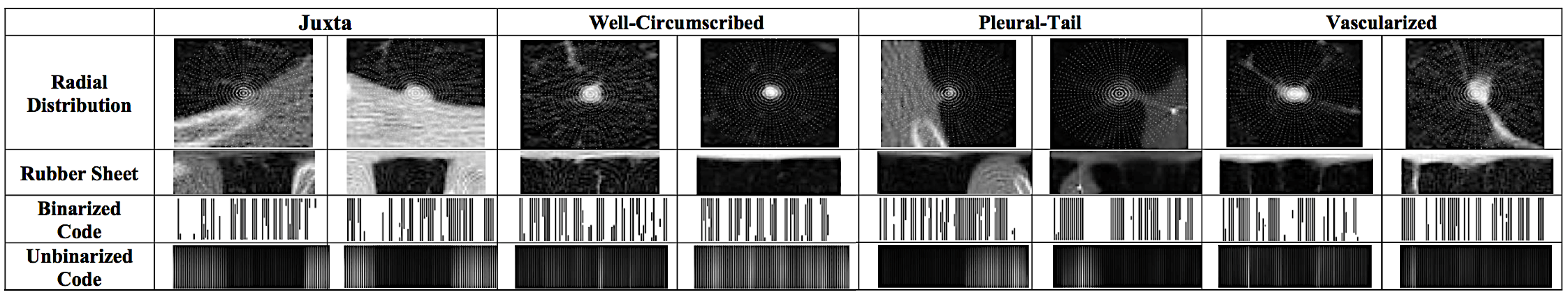
Visualization of Daugman recognition process for different nodule
types.
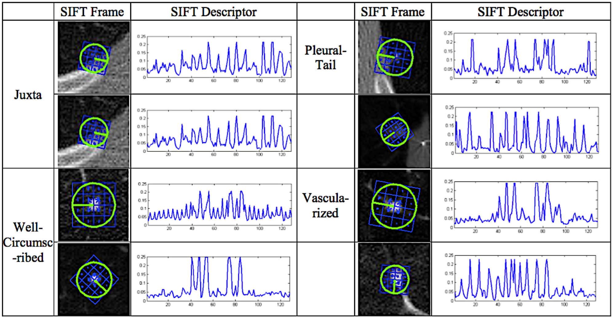
Visualization of SIFT recognition process for different nodule
types.
The Speeded Up Robust Features (SURF) and the Local Binary Pattern (LBP) descriptors were also used to generate the features that describe the texture of common lung nodules. These features were optimized and the resultant set was used for classification of lung nodules into four categories: juxta-pleural, well-circumscribed, vascularized and pleural-tail, based on the extracted information. Experimental results illustrated the efficiency of using multi-resolution feature descriptors, such as the SURF and LBP algorithms, in lung nodule classification.
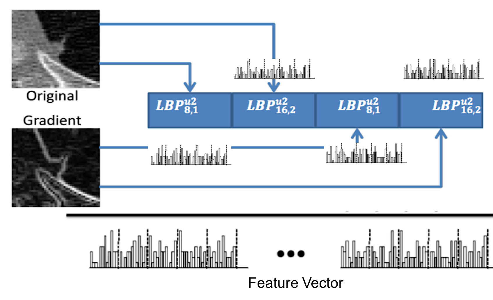
LBP feature vector generation using original and gradient nodule
images.
Related publications:
Amal Farag, Asem Ali, Shireen Y. Elhabian, James Graham, Aly Farag, Robert Falk. Feature-Based Lung Nodule Classification. Proc. of International Symposium on Visual Computing (ISVC), pp. 79-88, 2010.
Amal Farag, Shireen Y. Elhabian, James Graham, Aly Farag, Robert Falk. Toward Precise Pulmonary Nodule Descriptors for Nodule Type Classification. Proc. of the 13th International Conference on Medical Image Computing and Computer Assisted Intervention (MICCAI), pp. 626-633, 2010.
Modeling corpus callosum shape for early detection of autism
Joint work with: Aly Farag
Autism has no current cure or proven mechanism as to how it develops. Nonetheless, accompanying severity can be significantly reduced with the proper therapy, and thus the earlier that the disease is detected the faster therapy can be administered. This research is an attempt at studying discriminatory shape measures of related brain structures (in specific the corpus callosum) that are known to carry changes from autistics to normal individuals.
The Bezier curve and surface were used as parametric polynomial representations to model the shape of the Corpus Callosum (CC) region from T1-weighted clinical MRI scans. We drived a closed form solution for the Bezier coefficients in 2D and 3D Euclidean spaces. The coefficients of the models were used for reconstruction of the CC contours and surfaces with varying degrees of accuracy, and constituted basis for discrimination between populations, and ways to enhance elastic registration of the CC. The discrimination ability of the Bezier curves and surfaces were evaluated against the Fourier Descriptors (FD) and Spherical Harmonics (SH) approaches.
This approach was tested on T1-weighted MRI scans of 16 normal and 22 autistic subjects and showed promsing classification results, suggesting that this approach is worth investigating on a larger population with the hope of providing early identification and intervention of autism using neuroimaging.
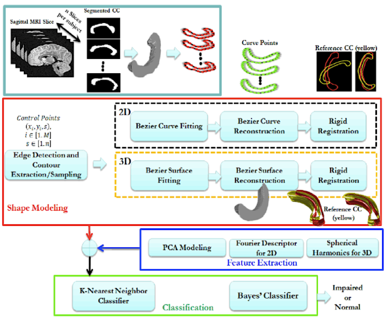
A block diagram of the proposed analysis system of the corpus callosum (CC) from T1-weighted MRI scans. From segmented MRIs, contours and surfaces of the CC structure are modeled using various parametric methods. Features are extracted and optimized to suite various classification approaches to distinguish between normal and abnormal CC.
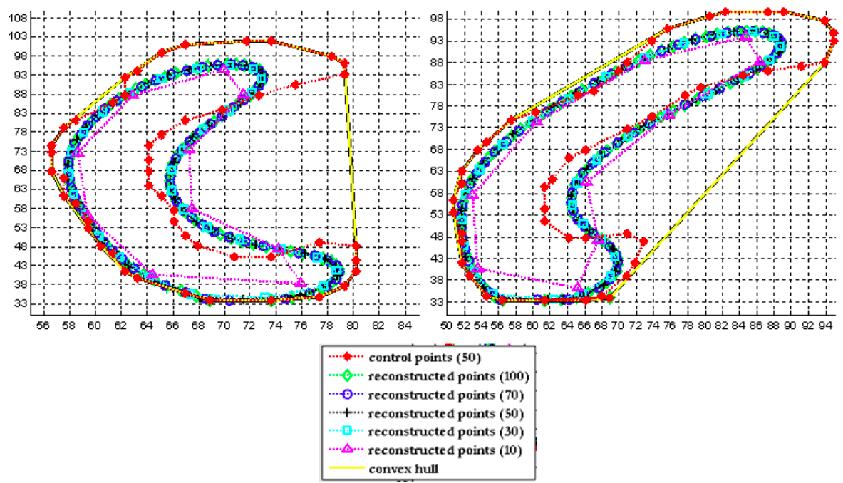
Bezier curve of corpus callosum for autistics (left) and normal subject (right). Control points are shown in red, and the convex hull of the control points is shown in dashed-yellow line. The reconstructed points are obtained by sampling the parameter \(t\in[0,1]\) at different resolutions; it is observed that the reconstructed curves are maintained within the convex hull of the control points.
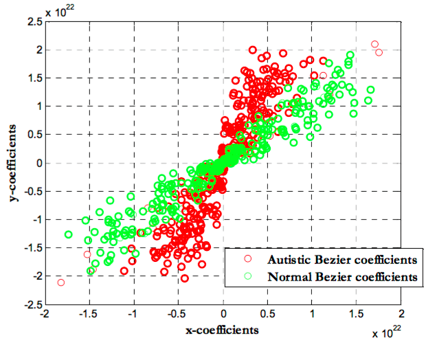
Scatter plot of the Bezier coefficients of the corpus callosum of
autistic and normal subjects.
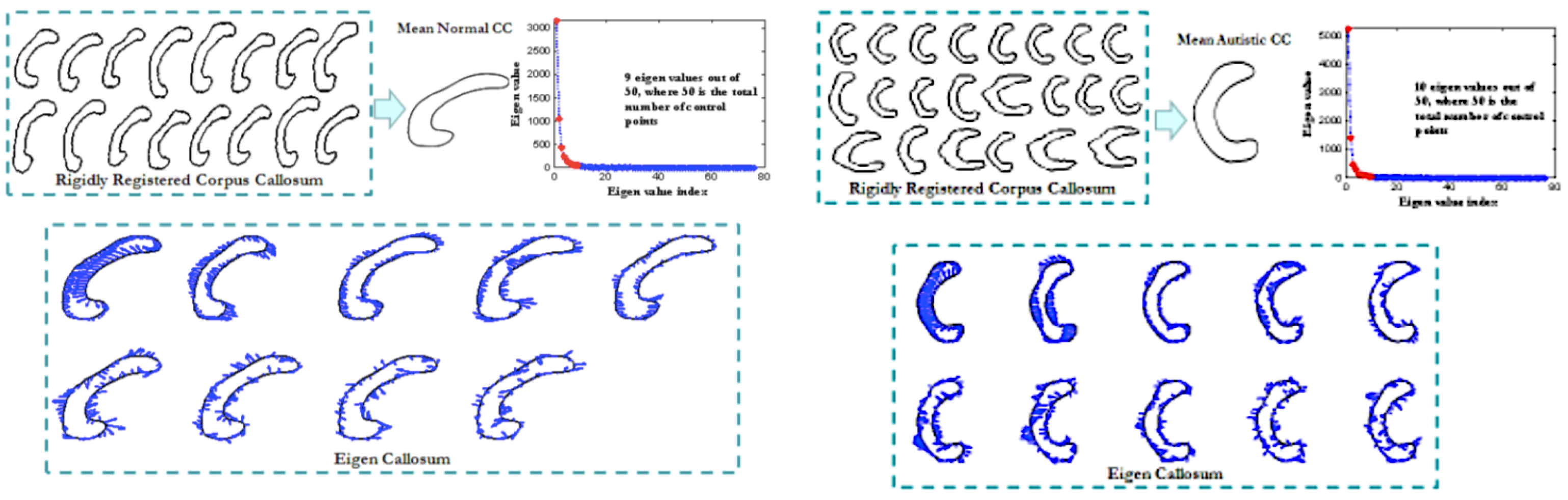
Given a set of training normal/autistic corpus callosum rigidly registered, a mean autistic CC is computed. The eigen-values spectrum of the covariance matrix of the zero-mean autistic CC is shown where only 9 (for normals) and 10 (for autistics) of them maintain 98% variability of the training data, eigen callosum which are the most significant eigen vectors of the covariance matrix are plotted (as directions of variations in xy-plane) with the mean autistic CC superimposed for visualization.
Related publications:
Ahmed Farag, Shireen Y. Elhabian, Mostafa Abdelrahman, James Graham, Aly Farag, Dongqing Chen, Manuel F. Casanova. Surface Modeling of the Corpus Callosum from MRI Scans. Proc. of International Symposium on Visual Computing (ISVC), pp. 9-18, 2010.
Ahmed Farag, Shireen Y. Elhabian, Mostafa Abdelrahman, James Graham, Aly Farag, Dongqing Chen, Manuel F. Casanova. Shape Modeling of the Corpus Callosum. Proc. of the 32nd IEEE Engineering in Medicine and Biology Society (EMBC), pp. 4288-4291, 2010.
Copyright © 2022 Shireen Y. Elhabian. All rights reserved.