Robert S. MacLeod, Quan Ni, Bonnie Punske, Philip R. Ershler, Bulent Yilmaz, and Bruno Taccardi
Presented at the 2000 International Society for Computerized Electrocardiography Conference, Yosemite, CA.
Previous studies have examined the influence of body position, respiration, and habitus on body surface potentials, however, the authors could only estimate the sources of the effects they documented. Among the proposed origin of changes in body surface potentials from those studies were the position of the heart, alterations in autonomic tone, differences in ventricular blood volume, and variations in torso resistivity. The goal of this study was to investigate specifically the role of geometric factors in altering body surface potentials and the ECG. For this, we used experiments with an isolated, perfused dog heart suspended in a realistically shaped electrolytic torso tank. The experimental preparation allowed us to measure epicardial and tank surface potentials simultaneously, and then reconstruct the geometry of both surfaces. Our results mimicked some of the features described by previous investigators. However, our results also showed differences that included considerably larger changes in the peak QRS and T-wave amplitudes with heart movement than those reported in human studies. We detected smaller values of root-mean-squared variability from heart movements than those reported in a human study comparing body surface potentials during change in inspiration and body position. There was better agreement with relative variability, which in these studies ranged from 0.11-0.42, agreeing well with an estimate from human studies of 0.40. Our results suggest that the isolated heart/torso tank preparation is a valuable tool for investigating the effects of geometric variation. Furthermore, the geometric position of the heart appears to be a large source of variation in body surface potentials. The size of these variations easily exceeded thresholds used to distinguish pathologic conditions and thus such variations could have important implications on the interpretation of the standard ECG.
The goal of the research presented here was to address a topic that has appeared regularly in the literature for at least the past sixty years[1,2,3,4,5,6]. Despite such extensive studies, there remain questions regarding the effect of heart position, body position, and respiratory status on the body-surface electrocardiogram. The technical scope of these studies spans the modern era of electrocardiography and most recently their authors have based their conclusions on large scale body surface potential mapping of human subjects[5,6]. A major limitation of all these studies, however, is the fact that the underlying mechanisms of variation remained inaccessible to direct observation.
In these studies, we have attempted to provide a more detailed and intimate examination of the mechanisms for electrocardiographic changes associated with heart location. We have utilized measurements from an instrumented, isolated heart preparation suspended inside a torso shaped electrolytic tank to assess directly the role of heart position--and indirectly that of body posture--on body surface potentials. This particular experimental preparation provides extensive access to and control of relevant parameters as well as the option of both qualitative and quantitative evaluations of the associated changes in the ECGs over the entire torso tank surface.
We performed a series of tests using the torso tank with the specific goal of comparing results in more simplified, controlled conditions than those described in two major studies of heart position and geometry. We sought to mimic the conditions described by Sutherland et al.[5] of placing the subjects in different body positions by moving the heart within the tank. We also used similar methods as Sutherland et al. for preprocessing and evaluating the resulting body surface maps recorded from the surface of the torso shape electrolytic tank and computed similar statistics. We borrowed heavily from the recent PhD thesis of Hoekema[14] to set the range of heart positions within the tank to match those that he described based on magnetic resonance images of human subjects. Furthermore, we evaluated a finding from his research that the statistical variability caused by geometric error was approximately 0.4. Hoekema was not able to measure separate variability values for geometric and physiologic influences but proposed a means of estimating these values based on measures of total variation and simulations using an electrocardiographic inverse solution. We were able to vary geometric and physiologic conditions independently and thus measure the geometric variability.
In general, the results from these experimental studies matched those published from studies in humans. The advantage of the experimental preparation is that the source of variation was unequivocally either geometry or physiological changes that we induced. Hence, it is possible to address in a controlled manner a large number of the factors that affect the relationship between electrical activity in the heart and its reflection on the body surface.
The preparation for the experiments was one we have used extensively for
the past five years at the CVRTI[7,8,9] and
consisted of an isolated dog heart, perfused by a second support dog and
suspended in an instrumented electrolytic tank. The tank had the shape of
an adolescent thorax and contained 370 Ag/AgCl pellets embedded in the
inside of the shell to detect tank surface potentials. 128 silver wires
sewn into a nylon stocking that was slipped over the ventricles measured
epicardial potentials from the isolated heart. Sampling rates for both
heart and tank recording were 1000 samples/s and a Wilson central terminal
served as reference for the unipolar electrograms and ECGs. To place an
appropriate electric load on the isolated heart, the electrolyte was a
mixture of isotonic saline and sucrose with a bulk resistivity of
500 ![]() -cm, similar to that of the human
thorax[10,11].
-cm, similar to that of the human
thorax[10,11].
To acquire all signals simultaneously, we have developed custom acquisition systems capable of recording from up to 1024 channels at 1000 samples/s and transferring the data directly to the magnetic disk of a Macintosh personal computer. Once saved, the data were available for immediate review by the acquisition system and then transfered to Unix workstations (Silicon Graphics, Inc.) for subsequent analysis, visualization, and archiving. Standard preprocessing of the cardiac electrograms and tank ECGs consisted of gain adjusting each input signal, adjusting baselines, and selecting a representative beat for each recording burst, either through windowing or time aligned signal averaging. From a root-mean-squared (RMS) curve for all the signals on each surface, we manually set fiducial markers for the onset and offset of the QRS complex and the end of the T wave. These fiducial markers provided the information necessary to calculate time integrals for various segments of each ECG and electrogram. Many of the results presented here are based on the QRS, STT, and QRST integrals and on the peak values of QRS and T-wave amplitudes.
To create physiological variation within the experiments, we paced the isolated heart from an atrial hook electrode and from bipole pairs located in the subepicardial regions of the right ventricle, anterior left ventricle, left ventricular free wall, left ventricular posterior wall, and the apex.
An important component of some experiments was the ability to move the heart in three orthogonal directions within the torso tank and to reconstruct the resulting geometry of heart electrode locations relative to the tank. This required the use of a gantry and clamp mechanism that held the heart in place. A mechanical digitizer (Microscribe) driven by custom software provided a means of tracking the location of the heart within the tank, as well as reference points from the tank surface. For each new heart location, we digitized 3-6 common reference points on the heart or the heart clamp to ensure proper location. At the end of the experiment, after fibrillating the heart to make it slightly more rigid but maintain it in a diastolic position, we raised the heart from the tank and digitized all 128 electrode locations, as well as the reference points, and the major branches of the coronary arteries (to use for visual references in subsequent visualizations). Repeated digitizations of the same hearts by the same and different observers in past experiments have indicated average errors of approximately 2-3 mm due to operator errors and heart movement (unpublished results).
When comparing potential distributions for different pacing sites or heart locations, we first examined the potentials as three-dimensional color-coded displays using our custom visualization software[12,13]. To quantify the differences between sets of the torso tank and epicardial potentials, we compared the values of parameters extracted from the maps, including peak values of the QRS and STT segment and QRS, QRST, and STT integrals. For statistical comparisons, we used the following metrics:
RMS =  , ,
|
(1) |
RelVar =  , ,
|
(2) |
Based on the results of Hoekema[14], and limited by the physical constraints of the torso tank and the gantry used to suspend the heart, we shifted the position of the heart in steps of 1 cm over all three orthogonal directions through the following ranges:
| Direction | Range [cm] |
| X-axis (left to right) | 6 |
| Y-axis (front to back) | 5 |
| Z-axis (up and down) | 5 |
Thus, with these variations of pacing site and heart location, the resulting data set can be pictured as a three-dimensional matrix with axes indicating variation in pacing site (atrial and five ventricular sites), shift direction (x, y, and z), and amount of shift (up to 6 cm), respectively. Each node of the matrix then contains a single heart beat, which we examined as a sequence of potential maps or in the form of extracted parameters such as peak values and integrals. Subsequent plots of these parameters (eg., Figures 3 and 4) show the variations of that parameter as a function of location along one of the axes of this data matrix.
Some features of the potential distribution on the tank surface changed as expected while other features showed unexpected results. Features such a locations of extrema and lines of zero amplitude shifted in the same direction as the shift in heart position. Figure 1 contains an example of a sequence of QRS integral maps recorded during a sequence of shifts of the heart from the right to left side in the torso tank with right ventricular pacing. The pattern of contour lines on the anterior view of the torso tank shifted along with the heart position, from the right towards the left side of the torso. The epicardial distributions, in contrast, were very stable throughout the sequence. The extrema of the tank-surface integral map distributions did not shift substantially in location but instead grew in amplitude as the heart moved from right to left. This is an expected result for the maximum because the underlying cardiac source moved closer to the left side of the tank and thus one would expect it to cause an increase in amplitude on the tank surface. The negative extremum on the right side of the tank, however, also increased slightly in amplitude and greatly in size with movement of the heart away from the right side of the tank, not an expected result. The finding that the epicardial potential distributions remained consistent throughout the shift in heart position suggests that physiologic variation was not likely to cause the changes in tank surface patterns.
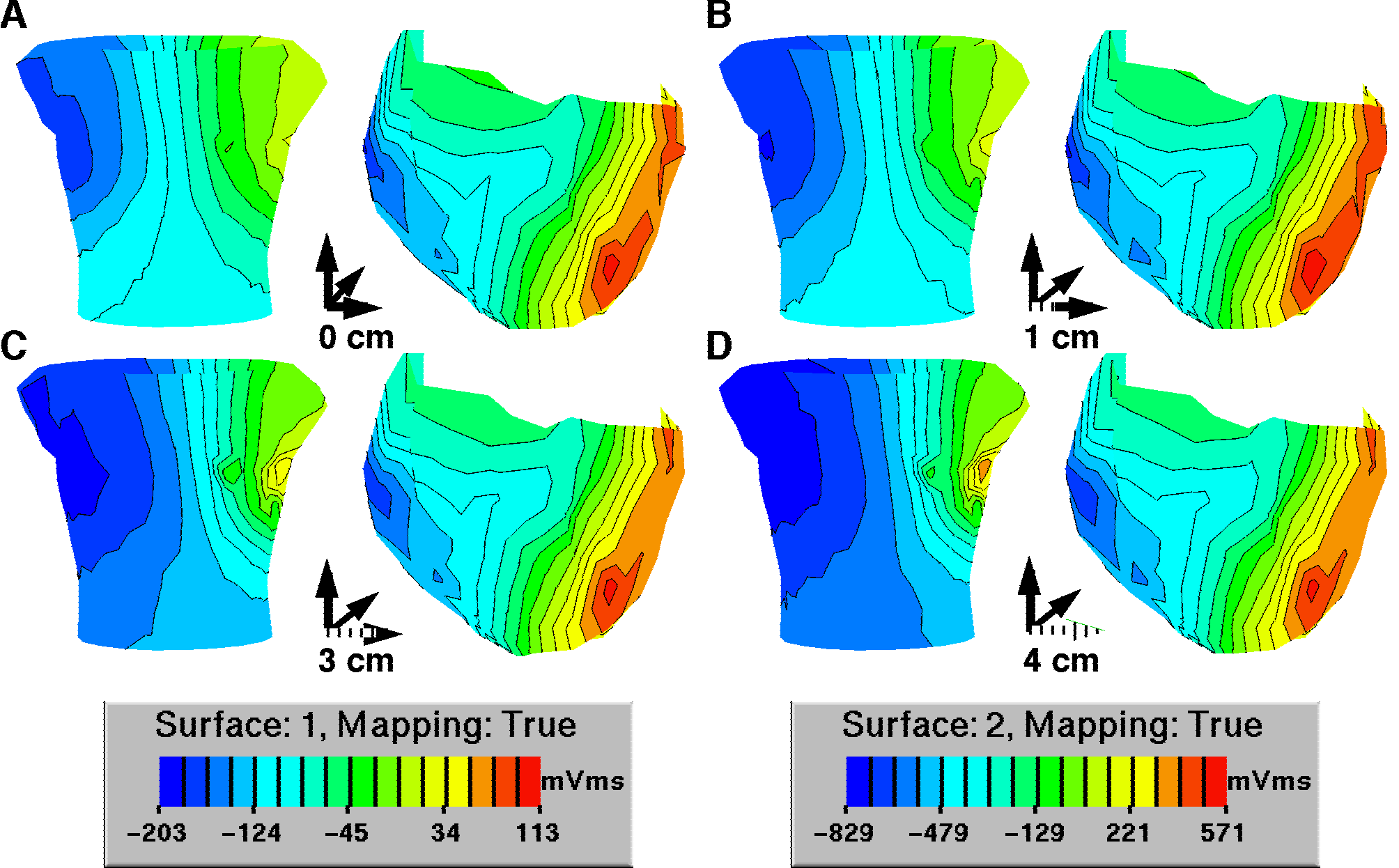 |
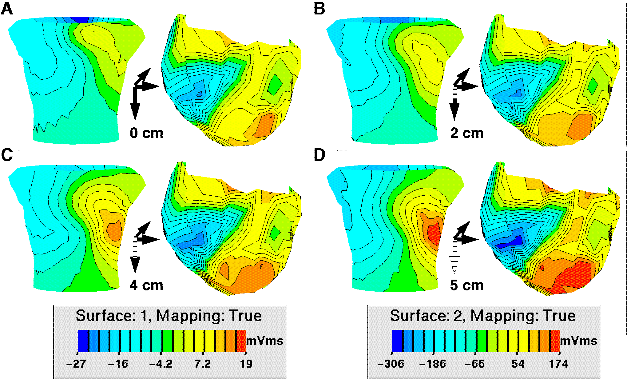
Figure 2 shows a sequence of QRS integral maps following movement of the heart from the top of the tank downward over 5 cm. Here too, there is the expected downward shift in the integral distribution, especially the positive values, but also a small rotation of the pattern and a growth in the maximum amplitude with downward shift. In this case, there was also a slight change in epicardial potential amplitude but relatively far smaller than the change in torso potential amplitude.
 |
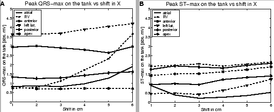
Sutherland et al. examined the change in peak amplitudes of the QRS and STT waves of the body surface ECG over all 120 leads with variation in body position (and, by extension, with variations in heart position within the body). We carried out a similar analysis for movement of the heart in the tank, one example of which is shown in Figure 3. This figure shows the variation in peak positive values in the QRS complex and STT segment as a function of shift in the x direction. These graphs revealed a wide variation in response with pacing site, which was repeated across all other movement directions. Some peak values underwent large rises--four- and five-fold in some cases--while others dropped, and still others remained constant. This variation in response was also visible in the sequences of tank surface maps in which extrema would rise, fall, or remain constant depending on the location of the associated feature on the heart relative to the tank surface and the direction of heart shift. For example, an anterior maximum might rise and a posterior minimum fall as the heart approached the front of the tank. However both might change in more complex ways as the heart moved from left to right at the same distance from the front surface of the tank, as shown in Figure 1, in which both extrema rose in amplitude with shift in heart location from right to left.
 |
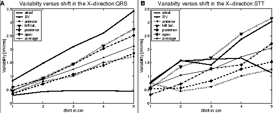
The ``variability index'' described above is one means of measuring the statistical changes in torso tank potentials induced by shifting the heart location within the tank. Sutherland et al. used this parameter to evaluate the extent of difference between integral maps in subjects in different postures and levels of inhalation[5].
Figure 4 A contains plots of the variability index of the QRS integral over the torso tank surface as a function of heart location shift in the x-direction. Each curve represents the sequence of variability index values as the heart moved from the right toward the left arm for a different pacing site. There was only slight change in the variability for atrial pacing but all other pacing sites showed a monotonic increase, in some cases over a factor of three, for a 5 cm of shift in heart location. Figure 4 B shows results from the same beats but for the STT integral. In this case, the atrial beats also showed substantial changes but with a plateau between 2 and 4 cm and a return to almost control at 5 cm. The other pacing sites produced generally increasing values of variability but the sequences were not always monotonic (eg., for anterior and right ventricular pacing.)
 |
Variability values for shifts in the y-direction (front to back) were generally smaller than those for the other two directions of shift. There was a similar monotonic trend to larger values of QRS-integral variability with shift, but the largest amplitudes of variability exceeded 1.0 in only a few cases. The pattern for variability of the STT integral with shift in the y-direction was similar to that for x-axis shift with a general but not monotonic increase with heart shift from the front towards the back of the tank. Variability values following shifts in the z-direction were similar to those with shift in the x-direction and of comparable magnitudes.
To summarize these results, Table 1 contains the ranges of variability indices for the different integrals and movement directions.
To capture the total variance in the tank potentials with changes in heart location, we computed the relative variance of the QRS integral for all beats from the same pacing site over all locations. The results for all pacing sites are summarized in Table 2
|
The strength of this study based on an experimental preparation compared to previous studies from human subjects lies in the ability to control both physiologic and geometric sources of variation independently in a reproducible manner. In these experiments, we varied the physiological conditions through choice of pacing site for the isolated heart and the geometric conditions by shifting the position of the heart within the tank. The main goal of this particular study was to compare our results with those from previous human studies as a means of evaluating the ability of the preparation to mimic the much more complex conditions of an intact human thorax.
In their examination of variability within a population of 36 normal subjects, Sutherland et al. computed variability indices using the same approach as we have presented[5]. The changes in subject conditions included in the Sutherland et al. study were in the level of respiration (tidal respiration, end-tidal inspiration, functional residual capacity, total lung volume, residual volume, and Valsalva and Müller maneuvers) and body position (standing versus supine) and the variability index used the standing/tidal respiration values as the reference. They found variability values that ranged from 2-7 mVms in the QRS integral, whereas the maximum variability in the torso tank ranged from 0.33-3.4 mVms for shifts of 5-6 cm, depending on the pacing site. Their results for the STT integral in the human subjects were considerably smaller, from approximately 1.0-3.5 mVms, while in the torso tank the range was similar to that for the QRS integrals: 0.74-3.1 mVms.
Sutherland et al. did not collect image data on the subjects of their study so it is difficult to speculate on the range of anatomical differences within their group. The fact that we found similar levels of variability in the isolated heart/torso tank preparation does, however, suggest that most of the variability measured in their patient group was probably due to geometric factors and not physiologic variation. This proposition is further supported by their finding that the largest variability values arose through changes in body position and the most dramatic changes in lung volume, all of which would lead to large variations in geometry. The similarity of results also shows the ability of the torso tank preparation to generate relevant results, but with a significantly higher level of control. Direct access to the heart provided not only the means to set its location, but also to measure, for example, potentials on both the torso tank and the epicardial surface and to calculate the associated variability index.
Hoekema used the relative variance as a measure of interindividual variation across all leads for 100 ms of the QRS over all 25 subjects in his study and produced a value of 0.519±0.002 [14]. The sources of this variability were twofold: a) physiologic variation between the beats from different individuals and different times; and b) variation due to the different geometries of each subject, which Hoekema also documented extensively. Hoekema went on to estimate the variation due to geometry based on inverse computations on standardized torso geometries as approximately 0.40 of the total, so that variability due to physiology was approximately 0.33 (relative variances add quadratically)[14].
In this study, we evaluated the relative variance across all heart locations and the QRS integral for each of the different pacing sites individually. This approach served to minimize physiologic variability (pacing site was constant) while inducing geometrical variation over the range of approximately 5-6 cm in each of the orthogonal directions. These values for geometric shift were similar to the range Hoekema observed from the magnetic resonance imaging based reconstructions of the subject geometries in his study. The resulting relative variance values in the experiments ranged from 0.11-0.42, in the same range as that estimate by Hoekema.
This finding not only supports the conclusions of Hoekema, but conversely shows that the isolated heart/torso tank preparation can reproduce results that are quite consistent with those from human subjects. The similarity with results from Sutherland et al. further supports the conclusion that the isolated heart/torso tank preparation is a very viable preparation for studies of geometric variations and their effect on the ECG. Moreover, the higher level of control possible in the experimental preparation permits clearer separation of geometric and physiologic influences. For this study, we carried out only shifts in heart location, while human hearts are often rotated at different angles as well as having different sizes, variations that are all possible with the isolated heart preparation.
Further notable findings of this study include that changes in ECG potentials with heart geometry are complex and do not follow a simple set of rules. The response to shift in heart position depends on the relationship among pacing sequence, heart orientation, and direction of shift. A further finding of clinical relevance is that the size of the changes in body surface potential distributions and amplitudes are large enough to be of concern for clinical diagnoses. Peaks in QRS amplitude varied over threefold in some cases, while STT peak potentials both rose and fell by factors of over two, depending on the pacing site.
Future studies based on this preparation will include performing rotational changes in heart location to mimic those described by Hoekema as well as the quantitative examination of the effects of physiological variation on variability indices. These studies will also lend themselves well to validation with computational simulations and the use of various compensation schemes to attempt to remove the effects of interindividual variability in torso geometry. One means of achieving such compensation would be to compute inverse solutions based on individualized torso geometries and thus generate the associated sources for each measured body surface map. One could then either compare these sources across individuals and develop diagnostic criteria or then compute some form of normalized body surface potentials based on a standard torso geometry. Hoekema described such an approach[14] and attempted to implement it, but was not as successful as anticipated. The isolated heart/torso tank preparation might serve as an excellent basis for developing and refining such schemes under much more controlled conditions than human studies would permit.
The authors wish to express their gratitude to Yonild Lian and Jayne Davis for expert technical assistance in the experimental studies, to Ted Dustman for development of analysis software, and to Bruce Steadman for the acquisition hardware.
Support for this research as come from awards from the Nora Eccles Treadwell Foundation and the Richard A. and Nora Eccles Harrison Fund for Cardiovascular Research, the Whitaker Foundation and the NIH SCOR grant in Sudden Cardiac Death, HL 52338-05.
This document was generated using the LaTeX2HTML translator Version 99.2beta6 (1.42)
Copyright © 1993, 1994, 1995, 1996,
Nikos Drakos,
Computer Based Learning Unit, University of Leeds.
Copyright © 1997, 1998, 1999,
Ross Moore,
Mathematics Department, Macquarie University, Sydney.
The command line arguments were:
latex2html -split 3 -no_white -link 3 -no_navigation -nomath -html_version 3.2,math paper
The translation was initiated by Rob MacLeod on 2000-11-09