 Congratulations to Yong Wan and Charles Hansen, et al on their winning submission to this year's FASEB BioArt Competition
A. Kelsey Lewis1, Yong Wan1, Mary Colasanto1, Mark van Langeveld1, Ronen Schweitzer2§, Charles D. Hansen1, and Gabrielle Kardon1§
1University of Utah, Salt Lake City, UT, 2Shriners Hospital, Portland, OR, §Society for Developmental Biology
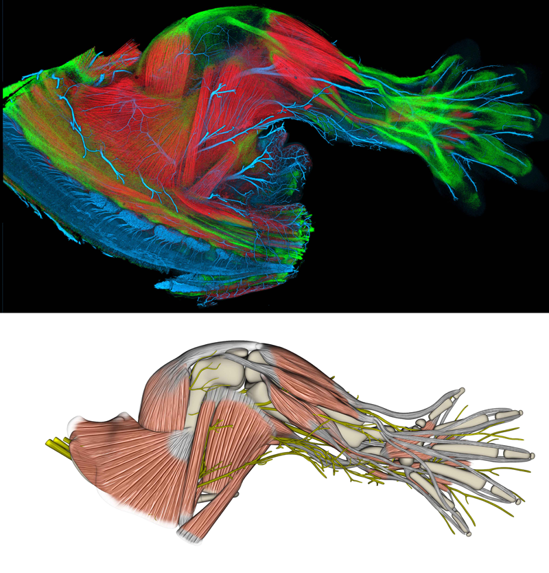
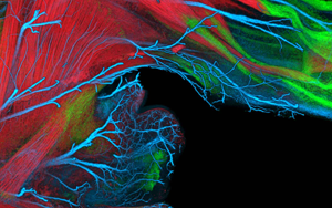
Upper panel:
This is a limb from a transgenic, embryonic mouse, showing well established musculoskeletal and nervous systems. The limb is stained with a variety of techniques to differentiate muscle, tendon, bone, and nerve, and rendered into a three-dimensional image using FluoRender, a publicly available rendering program developed at the University of Utah.
Lower panel:
This is a model, derived from the upper panel image, showing muscles, tendons, bones, and nerves. This model is part of a collaborative effort between geneticists and computer scientists to develop a 3-dimensional interactive atlas of limb development. NIH funding from the Eunice Kennedy Shriver National Institute of Child Health and Human Development supports this effort.