Statistical and Biomechanical Analysis of Hip Dysplasia
Collaborating Investigators: Dr. Jeffrey Weiss, Dr. Andrew AndersonAcetabular dysplasia is one of the leading causes of premature osteoarthritis (OA) of the hip. However, the relationship between the altered geometry associated with dysplasia and the resulting stresses in and around the joint is poorly understood. Recognizing the mechanical consequences of different and often subtle forms of dysplasia allows earlier identification of 'at risk' hips, in turn allowing the initiation of earlier treatment. This research delineates the true spectrum of this three-dimensional pathology by quantifying stress transfer in the hip joint and predicting the long-term success rate of corrective surgeries.
Much of orthopaedics is governed by mechanics and much of mechanics is dictated by shape. Thus, we believe that the potential impact of robust, open-source software tools for meshing and statistical shape analysis is profound. The ability to study statistical shape models in a biomechanical context has several important implications, including the ability to drive the biomechanical simulations of patient groups using group-averaged geometries, the ability to make group comparisons of both geometry and biomechanical parameters in a way that systematically captures group variability, and finally, the ability to study sensitivities in biomechanical outcomes with respect to geometric variability. This project presents some important challenges for algorithm and software development in the CIBC: (1) achieving a sufficient level of robustness to compete with the commercial pipeline; (2) validating the quality of the elements in the biomechanics applications; (3) extending the tools in a general way to deal with open geometries; (4) addressing some of the issues of building joint models (multiple surfaces) with tight tolerances (bones separated by cartilage); and (5) achieving a proper anatomical alignment of the bones.
Statistical Shape Modeling of Cam Femoroacetabular Impingement
Cam femoroacetabular impingement (FAI) is characterized by a malformed femoral head that may cause shearing between the femur and acetabulum, leading to intraarticular damage and early hip osteoarthritis. Radiographic measurements are used to diagnose cam FAI, but provide only a planar view of the femoral head and often assume the ideal femur shape to be spherical. Statistical shape modeling (SSM) can be used to objectively compare complex 3D morphology without the need to assume ideal geometry. The objective of this study was to generate accurate 3D reconstructions of femoral heads and apply statistical shape modeling to quantify 3D variation and morphologic differences between control and cam femurs. Femurs from 33 controls and 15 cam FAI patients were CT scanned and 3D surfaces were generated by image segmentation. Correspondence particles were optimally positioned upon each surface using a gradient descent energy function. Resulting particle configurations were used to generate mean shapes for each group. Morphological differences were calculated as the distance between mean control and patient geometries. Differences were consistent with the location and approximate shape of cam lesions found intraoperatively. Deviations in mean shape between groups were pronounced at the anterolateral head-neck junction, where the mean cam femur protruded from the mean control femur by a maximum of 2.7mm. Sustained protrusions of ~1.0--2.5mm were distributed from the anterior-posterior midline of the femoral neck along the entire anterolateral head-neck junction and distally along the anterior section of the neck. Future work will refine our statistical shape modeling software to quantify, on a patient-specific basis, the severity of cam lesions for preoperative planning. This project was accepted for publication in Journal of Orthopedic Research.This work was published as an abstract and was presented at the Orthopedic Research Society 2013 Annual Meeting (ORS 2013) on Statistical Shape Modeling of Cam Femoroacetabular Impingement1, and will appear as a journal article. Finally, a R21 NIH grant "Musculoskeletal and Finite Element Modeling of Femoroacetabular Impingement" was awarded to our collaborator from the Department of Orthopaedics, Andrew Anderson, PhD. This project, which grew directly from the CIBC collaboration, will start in June, 2013.
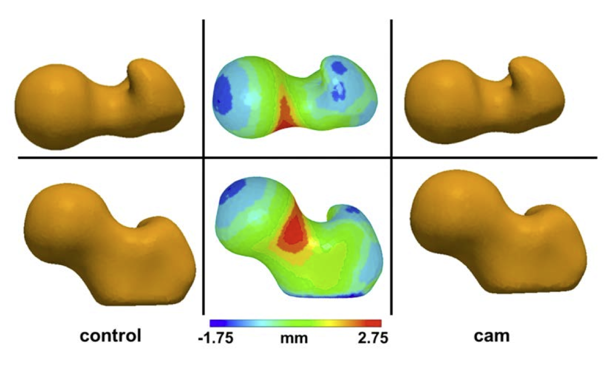 |
| Mean control (left) and cam (right) shapes. Middle images show the mean control shape with color plots depicting how the mean cam shape differed across the femoral head, neck, and proximal shaft. Top and bottom rows show different rotations of the femoral head. |