
 |
Ruth KlepferPrincipal Scientist at Medtronic Ph.D. in Bioengineering, 1997 Dissertation Title: Conductivity and Mesh Refinement in Finite Element Computations of Electrocardiographic Fields Advisor: Christopher R. Johnson |
Dr. Klepfer has been working as a scientist at Medtronic in Cardiac Rhythm and Disease Management Research for the past 12 years. In this role she is responsible for study design, protocol development, conduction of studies (both animal and clinical), anddata analysis and interpretation. She also collaborates with physicians in a consulting role and partners with physicians on various studies.
Dr. Klepfer's work focuses on potential therapies and diagnostics for people with congestive heart failure. For example, using coupled pacing or high-amplitude stimulation during the absolute refractory period as a means of increasing contractility in people with heart failure. Dr. Klepfer also, with a focus on heart failure, has studiedstimulation of the vagus nerve, pacing strategies for heart failure patients with a preserved ejection fraction, and the use of pulmonary pressure to better manage patients with heart failure.
Ruth received her Ph.D. in Bioengineering from the University of Utah in 1997. At the SCI Institute, Dr. Klepfer worked with Drs. Chris Johnson and Rob Macleod, and constructed a computer model of the macroscopic electrophysiology of the heart and torso using the finite element method (FEM). She quantified effects of three different sources of variability on simulated electrocardiographic (ECG) and implantable cardioverter defibrillator (ICD) fields. The geometry of the model was obtained from MRI's covering the entire torso with special attention given to anatomical and tissue detail near the heart. Her project involved extensive coding of the numerical analysis, finite element methods, data analysis techniques as well as an understanding of electrophysiology and cardiology. Previous to joining the SCI Institute, Ruth was a senior consultant at Accenture.
 |
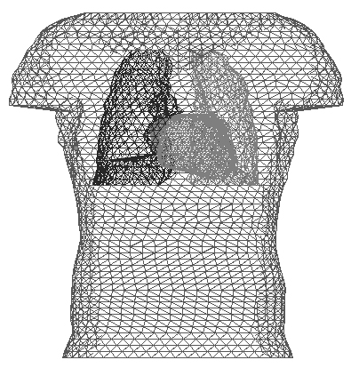 |
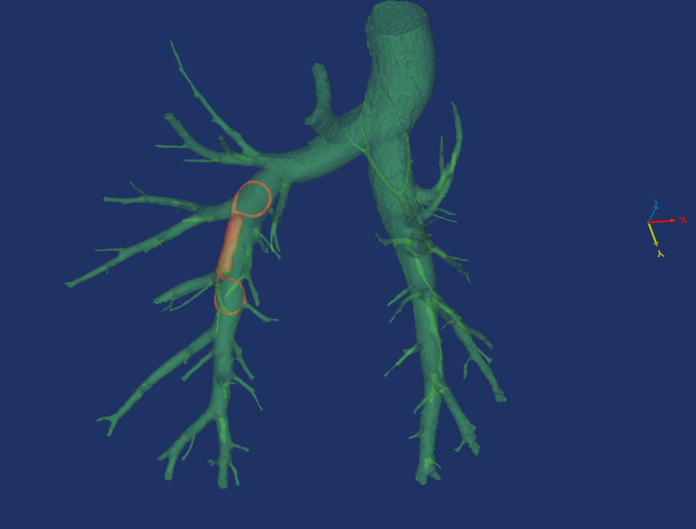
| At Medtronic - The figure shows a pressure sensor (red) implanted in the right pulmonary artery (green) of a sheep. These images are 3D objects reconstructed from CT scans taken of the animal just after implant. | At SCI - Frontal view of the surface triangulation of the Utah torso model. Surfaces included in the figure are torso boundaries, lungs and epicardium. |