Localization of Ischemic Change during PTCA
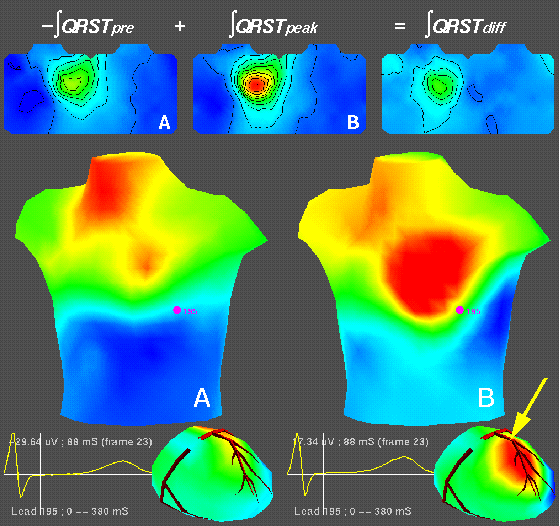
An example of how we detect changes due to ischemia in patients undergoing
coronary angioplasty. The top panel contains two isointegral maps from the
QRST segment, and their difference. The left map (A) originates from
signals measured before inflation of the PTCA balloon while the middle map
(B) is the same integral from a beat recorded at the latter, peak phase of
the inflation; the rightmost map is the difference (peak- minus
pre-inflation). These maps have been projected onto a two-dimensional
surface, with the anterior torso on the left of each map and the posterior
on the right. In the lower part of the figure, all distributions shown are
isopotential instant maps, also from the same pre-inflation (panel A) and
peak-inflation (Panel B) beats. The torso maps were measured, but the
epicardial maps in the lower right portion of each panel contain estimates
based on inverse calculations. The red areas denote positive potential,
the blue denote negative potential and the green/yellow colors span the
region of small potential values near zero. The arrow indicates the
approximate location of the angioplasty balloon. The presense of positive
potentials in the epicardial maps identifies the zone of predicted
ischemia, located downstream from the site of occlusion. This was work
from my PhD research performed with Milan Horacek at Dalhousie University.
Last modified: Wed Sep 11 19:43:43 MD 2002