Cardiac Mapping
Here are some examples of cardiac mapping, the high resolution measurement
of electrical activity from the heart.

Epicardial surface maps
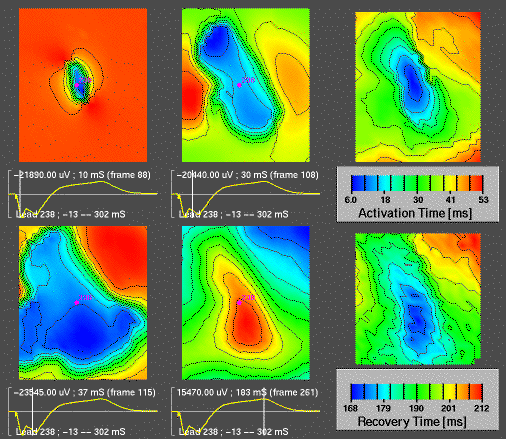
Isopotential and isochrone maps from a
regular mesh of electrodes sutured to the right ventricle of a heart. The
electrode array contained 21 x 25 electrodes regularly spaced at
2.0~mm separation and sewn into a nylon sock. Scaling---and thus contour
spacing---is local to each frame such that the colors span the range of
measured potentials. The 4 leftmost maps are isopotential instant maps from
the same heartbeat. In these maps the positive potentials are mapped to red
and negative potentials are mapped to blue. The time signal below each map
corresponds to the location in the maps marked by the red dot and indicates
how the maps progress through the beat. The rightmost column of maps were
derived from the entire beat and represent the activation times (upper map)
and recovery times (lower map) with scaling as indicated in the legends.
The data come from experiments carried out by Bruno Taccardi and Quan Ni
prepared the activation and recovery times.

Epicardial surface maps
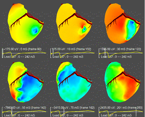
A sequence of isopotentials maps from an
sock electrode array containing 490 electrodes on semi-regular, curved grid
over the entire ventricles. Red outlines show the location of major
coronary arteries. Color coding for potentials is local to each instant
ranging from dark blue for most negative potentials, through green, yellow,
to red representing the most postive values. The time signal below each
map is marked by a vertical line to show progress through the beat. Bruno
Taccardi provided the surgical and experiment expertise.
Last modified: Wed Sep 11 19:45:50 MD 2002


