


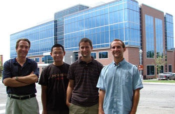
 (L-R) Dr. Guido Gerig, Dr. Marcel Prastawa, Casey Goodlett, Sylvain Gouttard |
Dr. Gerig is joined by three of his colleagues from UNC. Marcel Prastawa was a grad student under Dr. Gerig until he received his Ph.D. at the end of 2006. Since then he has worked as a Postdoc and now joins the SCI Institute as a Research Assistant Professor. Sylvain Gouttard has worked with Dr. Gerig for the past two years as a student intern from France. He developed software for the UNC's Neuroimaging Lab and assisted in using the software for applications such as clinical brain imaging studies. Sylvain will serve the SCI Institute as a research associate and will continue to develop tools for visualization and analysis of medical images as well as perform analysis and processing brain MRI images for research studies. Casey Goodlett joined Dr. Gerig as a Ph.D. student in the fall of 2004. He will continue pursuit of his Ph.D. as a graduate student at SCI working on diffusion tensor imaging (DTI) for the National Alliance for Medical Image Computing (NA-MIC).
Dr. Gerig will be the Institutes's first USTAR faculty member. The Utah Science, Technology and Research (USTAR) initiative is a state funded outreach and innovation program designed to promote local commercialization of discoveries and technologies emanating from the State's research universities and entrepreneurial activities. USTAR will fund a new Center for Neuroimage Analysis headed by Dr. Gerig that will collaborate as part of the University of Utah's Brain Institute. The Center will participate in the Brain Institute's mission to address basic research questions on brain function as a means for future improvement of treatment for neurological disorders and mental illness. The team will work closely with UCAIR, the Utah Center for Advanced Imaging Research, on questions of new state-of-the-art imaging sequences suitable to address relevant research questions. The Center will by definition be very closely related to the Scientific Computing and Imaging Institute since advanced high-performance computing, development of image processing tools, and scientific visualization are key elements for medical image analysis and large clinical studies.
Image Aquisition
The Neuroimage Analysis Center will work with radiologists and imaging researchers to determine optimal acquisition techniques for performing the CT and MRI scans for research studies. The current trend in imaging goes towards higher spatial resolution in three dimensions and acquisition of multiple contrasts. The scanning process often involves trade offs between scan speed and data quality. Longer scans can achieve higher resolution, however, however, scan time is limited considering patient comfort, costs of machine use, and minimal subject motion. The Center also helps to improve scan quality by developing methods for quality control, calibration of geometric and intensity distortion, and removal of imaging artifacts.Image Processing and Visualization
Once the data has been acquired from the scanner, Image Processing is required to extract the quantitative information from the images as requested by clinical researches. This might include brain volume, blood flow, size and shape of brain structures, thickness of cortex, or functional activation, for example. Maintaining software tools for various applications and developing advanced tools targeting new challenging problems are the primary focus areas of the Center. This includes training and teaching on methodology and supervision of trainees to get proficient on the use of methods. Image processing includes improving the contrast to noise ratio of the images, correcting for image distortion and artifacts, segmentation - separating the image into different types of anatomical and functional regions, and 3D modeling for morphometric analysis and scientific visualization. Designing easy to use, intuitive, GUI based tools that allow researchers to easily process and visualize the salient features of the data is also an important challenge.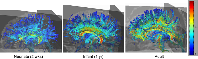 White matter fiber tracts via DT-MRI: Study of Early Development. Corpus callosum: Commissural bundles, color coding of fractional anisotropy FA (0=blue, 1=red) |
Feature and Statistical Analysis
Beyond aquisition and visualization of the data, it is also important to understand the meaning of the data. Feature analysis refers to the study of the condition of features based on morphology or other visible attributes. Often this involves comparing the size and shape of a feature to some standard and correlating that information with symptoms or clinical data taken from a patient. Measuring and describing the morphology of parts of the brain in a quantitative and meaningful way is an important challenge for the Center. The Center will also work in collaboration with neuroscientists and biostaticians to help test various hypotheses related to their research.Dr. Gerig and his team will also continue to work on four major multi-site projects begun at UNC.
- The Autism Center of Excellence (ACE).
- The National Alliance for Medical Image Computing (NA-MIC)
- The Biological Research Partnership (BRP)
- The CONTE Center for schizophrenia research.
The Brain Imaging Institute is currently recruiting faculty and collaborators for future work. As new talent is brought together, the path of future research will become clear.
Dr. Gerig expressed his thoughts about moving to Utah:
"There is a critical mass of researchers with excellence in various disciplines, and a very strong track record of multi-disciplinary collaborations so I was quite comfortable coming here. Many of them were already research colleagues on multi-center national projects and collaborators on various research projects."The SCI Institute is honored to have Dr. Gerig and his team join our ranks. We look forward with excitement at what the future holds.
"Utah is still the 'Wild West' in some respects. The ways of thinking and doing things are still being formed. There is a sense of pioneering the future and approaching problems in bold new ways. This attitude tends to spread to others who come here."
"I'm enthusiastic that my expertise in collaborative neuroimage analysis research coupled with the excellence of the SCI Institute and with the excitement of the University to form a strong Brain Institute will help us to make a difference and to have a strong impact." - Dr. Guido Gerig
For more information See:
Publications -- Guido Gerig