


SCI Publications
2013


J.M. Gililland, L.A. Anderson, H.B. Henninger, E.N. Kubiak, C.L. Peters.
“Biomechanical analysis of acetabular revision constructs: is pelvic discontinuity best treated with bicolumnar or traditional unicolumnar fixation?,” In Journal of Arthoplasty, Vol. 28, No. 1, pp. 178--186. 2013.
DOI: 10.1016/j.arth.2012.04.031
Pelvic discontinuity in revision total hip arthroplasty presents problems with component fixation and union. A construct was proposed based on bicolumnar fixation for transverse acetabular fractures. Each of 3 reconstructions was performed on 6 composite hemipelvises: (1) a cup-cage construct, (2) a posterior column plate construct, and (3) a bicolumnar construct (no. 2 plus an antegrade 4.5-mm anterior column screw). Bone-cup interface motions were measured, whereas cyclical loads were applied in both walking and descending stair simulations. The bicolumnar construct provided the most stable construct. Descending stair mode yielded more significant differences between constructs. The bicolumnar construct provided improved component stability. Placing an antegrade anterior column screw through a posterior approach is a novel method of providing anterior column support in this setting.
H.B. Henninger, C.J. Underwood, S.J. Romney, G.L. Davis, J.A. Weiss.
“Effect of Elastin Digestion on the Quasi-Static Tensile Response of Medial Collateral Ligament,” In Journal of Orthopaedic Research, pp. (published online). 2013.
DOI: 10.1002/jor.22352
Elastin is a structural protein that provides resilience to biological tissues. We examined the contributions of elastin to the quasi-static tensile response of porcine medial collateral ligament through targeted disruption of the elastin network with pancreatic elastase. Elastase concentration and treatment time were varied to determine a dose response. Whereas elastin content decreased with increasing elastase concentration and treatment time, the change in peak stress after cyclic loading reached a plateau above 1 U/ml elastase and 6 h treatment. For specimens treated with 2 U/ml elastase for 6 h, elastin content decreased approximately 35%. Mean peak tissue strain after cyclic loading (4.8%, p ≥ 0.300), modulus (275 MPa, p ≥ 0.114) and hysteresis (20%, p ≥ 0.553) were unaffected by elastase digestion, but stress decreased significantly after treatment (up to 2 MPa, p ≤ 0.049). Elastin degradation had no effect on failure properties, but tissue lengthened under the same pre-stress. Stiffness in the linear region was unaffected by elastase digestion, suggesting that enzyme treatment did not disrupt collagen. These results demonstrate that elastin primarily functions in the toe region of the stress–strain curve, yet contributes load support in the linear region. The increase in length after elastase digestion suggests that elastin may pre-stress and stabilize collagen crimp in ligaments
P.T. Scheffel, H.B. Henninger, R.T. Burks.
“Relationship of the intercondylar roof and the tibial footprint of the ACL: implications for ACL reconstruction,” In American Journal of Sports Medicine, Vol. 41, No. 2, pp. 396--401. 2013.
DOI: 10.1177/0363546512467955
Background: Debate exists on the proper relation of the anterior cruciate ligament (ACL) footprint with the intercondylar notch in anatomic ACL reconstructions. Patient-specific graft placement based on the inclination of the intercondylar roof has been proposed. The relationship between the intercondylar roof and native ACL footprint on the tibia has not previously been quantified.
Hypothesis: No statistical relationship exists between the intercondylar roof angle and the location of the native footprint of the ACL on the tibia.
Study Design: Case series; Level of evidence, 4.
Methods: Knees from 138 patients with both lateral radiographs and MRI, without a history of ligamentous injury or fracture, were reviewed to measure the intercondylar roof angle of the femur. Roof angles were measured on lateral radiographs. The MRI data of the same knees were analyzed to measure the position of the central tibial footprint of the ACL (cACL). The roof angle and tibial footprint were evaluated to determine if statistical relationships existed.
Results: Patients had a mean ± SD age of 40 ± 16 years. Average roof angle was 34.7° ± 5.2° (range, 23°-48°; 95% CI, 33.9°-35.5°), and it differed by sex but not by side (right/left). The cACL was 44.1% ± 3.4% (range, 36.1%-51.9%; 95% CI, 43.2%-45.0%) of the anteroposterior length of the tibia. There was only a weak correlation between the intercondylar roof angle and the cACL (R = 0.106). No significant differences arose between subpopulations of sex or side.
Conclusion: The tibial footprint of the ACL is located in a position on the tibia that is consistent and does not vary according to intercondylar roof angle. The cACL is consistently located between 43.2% and 45.0% of the anteroposterior length of the tibia. Intercondylar roof–based guidance may not predictably place a tibial tunnel in the native ACL footprint. Use of a generic ACL footprint to place a tibial tunnel during ACL reconstruction may be reliable in up to 95% of patients.
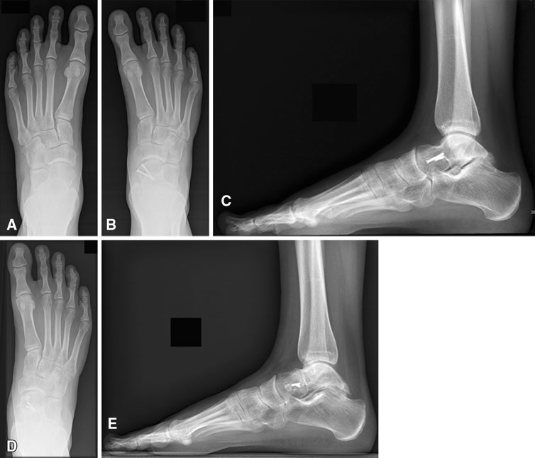
T. Suter, A. Barg, M. Knupp, H.B. Henninger, B. Hintermann.
“Surgical technique: talar neck osteotomy to lengthen the medial column after a malunited talar neck fracture,” In Clinical Orthopaedics & Related research, Vol. 471, No. 4, pp. 1356--1364. 2013.
DOI: 10.1080/10255842.2013.809711
PubMed ID: 23809004
R.Z. Tashjian, H.B. Henninger.
“Biomechanical evaluation of subpectoral biceps tenodesis: dual suture anchor versus interference screw fixation,” In Journal of Shoulder and Elbow Surgery, Vol. 22, No. 10, pp. 1408–-1412. 2013.
DOI: 10.1016/j.jse.2012.12.039
Background
Subpectoral biceps tenodesis has been reliably used to treat a variety of biceps tendon pathologies. Interference screws have been shown to have superior biomechanical properties compared to suture anchors; although, only single anchor constructs have been evaluated in the subpectoral region. The purpose of this study was to compare interference screw fixation with a suture anchor construct, using 2 anchors for a subpectoral tenodesis.
Methods
A subpectoral biceps tenodesis was performed using either an interference screw (8 × 12 mm; Arthrex) or 2 suture anchors (Mitek G4) with #2 FiberWire (Arthrex) in a Krackow and Bunnell configuration in seven pairs of human cadavers. The humerus was inverted in an Instron and the biceps tendon was loaded vertically. Displacement driven cyclic loading was performed followed by failure loading.
Results
Suture anchor constructs had lower stiffness upon initial loading (P = .013). After 100 cycles, the stiffness of the suture anchor construct "softened" (decreased 9%, P < .001), whereas the screw construct was unchanged (0.4%, P = .078). Suture anchors had significantly higher ultimate failure strain than the screws (P = .003), but ultimate failure loads were similar between constructs: 280 ± 95 N (screw) vs 310 ± 91 N (anchors) (P = .438).
Conclusion
The interference screw was significantly stiffer than the suture anchor construct. Ultimate failure loads were similar between constructs, unlike previous reports indicating interference screws had higher ultimate failure loads compared to suture anchors. Neither construct was superior with regards to stress; although, suture anchors could withstand greater elongation prior to failure.
2012
A. Barg, N. Knupp, H.B. Henninger, L. Zwicky, B. Hintermann.
“Total ankle replacement using HINTEGRA, an unconstrained, three-component system: surgical technique and pitfalls,” In Foot and Ankle Clinics, Vol. 17, No. 4, pp. 607--635. 2012.
DOI: 10.1016/j.fcl.2012.08.006
A. Barg, G.I. Pagenstert, A.G. Leumann, A.M. Müller, H.B. Henninger, V. Valderrabano.
“Treatment of the Arthritic Valgus Ankle,” In Foot and Ankle Clinics, Vol. 17, No. 4, pp. 647--663. 2012.
DOI: 10.1016/j.fcl.2012.08.007
A. Barg, M.D. Harris, H.B. Henninger, R.L. Amendola, C.L. Saltzman, B. Hintermann, A.E. Anderson.
“Medial distal tibial angle: comparison between weightbearing mortise view and hindfoot alignment view,” In Foot & Ankle International, Vol. 33, No. 8, pp. 655--661. 2012.
DOI: 10.3113/FAI.2012.0655
Background: The medial distal tibial angle (MDTA) is used to determine ankle alignment. The mortise view is the standard to measure MDTA, but the hindfoot alignment view (HAV) has become popular. The MDTA may vary between views, influencing the choice of surgery.
Methods: The MDTA was compared between the mortise and HAV in 146 ankles. MDTA was correlated to age and sagittal tibial tilt for each view. Differences in MDTA by gender and ethnicity were assessed. Diagnostic agreement (varus, valgus, normal) between views was calculated. Clinical assessment of alignment was determined and percent agreement between clinical and radiographic alignment was quantified.
Results: The MDTA measured from the mortise view and HAV radiographs was 89.0 (range, 81 to 96 degrees; SD = 2.8) degrees and 86.0 (range, 73 to 95 degrees; SD = 3.5) degrees, respectively. The MDTA was comparable for both genders for mortise (p = 0.356) and HAV (p = 0.621). The MDTA was comparable in all ethnic groups for mortise view (p = 0.616) and HAV (p = 0.916). Correlation between the measured MDTA and age was not statistically significant for both the mortise (r = 0.118; p = 0.158) and HAV (r = 0.148; p = 0.074). In only 47.3% of all ankles was the radiographic diagnosis of alignment the same between views. Agreement between clinical and radiographic classifications was 60.3% for the mortise view and 52.8% for the HAV.
Conclusion: Substantial disagreement in primary alignment was found between the mortise and HAV as quantified by the MDTA. Agreement between clinical and radiographic alignment was also poor. Clinical Relevance: Advanced imaging such as CT or MRI may better describe ankle alignment.
H.B. Henninger, Barg A, A.E. Anderson, K.N. Bachus, R.Z. Tashjian, R.T. Burks.
“Effect of deltoid tension and humeral version in reverse total shoulder arthroplasty: a biomechanical study,” In Journal of Shoulder and Elbow Surgery, Vol. 21, No. 4, pp. 483–-490. 2012.
DOI: 10.1016/j.jse.2011.01.040
Background
No clear recommendations exist regarding optimal humeral component version and deltoid tension in reverse total shoulder arthroplasty (TSA).
Materials and methods
A biomechanical shoulder simulator tested humeral versions (0°, 10°, 20° retroversion) and implant thicknesses (-3, 0, +3 mm from baseline) after reverse TSA in human cadavers. Abduction and external rotation ranges of motion as well as abduction and dislocation forces were quantified for native arms and arms implanted with 9 combinations of humeral version and implant thickness.
Results
Resting abduction angles increased significantly (up to 30°) after reverse TSA compared with native shoulders. With constant posterior cuff loads, native arms externally rotated 20°, whereas no external rotation occurred in implanted arms (20° net internal rotation). Humeral version did not affect rotational range of motion but did alter resting abduction. Abduction forces decreased 30% vs native shoulders but did not change when version or implant thickness was altered. Humeral center of rotation was shifted 17 mm medially and 12 mm inferiorly after implantation. The force required for lateral dislocation was 60% less than anterior and was not affected by implant thickness or version.
Conclusion
Reverse TSA reduced abduction forces compared with native shoulders and resulted in limited external rotation and abduction ranges of motion. Because abduction force was reduced for all implants, the choice of humeral version and implant thickness should focus on range of motion. Lateral dislocation forces were less than anterior forces; thus, levering and inferior/posterior impingement may be a more probable basis for dislocation (laterally) than anteriorly directed forces.
Keywords: Shoulder, reverse arthroplasty, deltoid tension, humeral version, biomechanical simulator
H.B. Henninger, A. Barg, A.E. Anderson, K.N. Bachus, R.T. Burks, R.Z. Tashjian.
“Effect of lateral offset center of rotation in reverse total shoulder arthroplasty: a biomechanical study,” In Journal of Shoulder and Elbow Surgery, Vol. 21, No. 9, pp. 1128--1135. 2012.
DOI: 10.1016/j.jse.2011.07.034
Background
Lateral offset center of rotation (COR) reduces the incidence of scapular notching and potentially increases external rotation range of motion (ROM) after reverse total shoulder arthroplasty (rTSA). The purpose of this study was to determine the biomechanical effects of changing COR on abduction and external rotation ROM, deltoid abduction force, and joint stability.
Materials and methods
A biomechanical shoulder simulator tested cadaveric shoulders before and after rTSA. Spacers shifted the COR laterally from baseline rTSA by 5, 10, and 15 mm. Outcome measures of resting abduction and external rotation ROM, and abduction and dislocation (lateral and anterior) forces were recorded.
Results
Resting abduction increased 20° vs native shoulders and was unaffected by COR lateralization. External rotation decreased after rTSA and was unaffected by COR lateralization. The deltoid force required for abduction significantly decreased 25% from native to baseline rTSA. COR lateralization progressively eliminated this mechanical advantage. Lateral dislocation required significantly less force than anterior dislocation after rTSA, and both dislocation forces increased with lateralization of the COR.
Conclusion
COR lateralization had no influence on ROM (adduction or external rotation) but significantly increased abduction and dislocation forces. This suggests the lower incidence of scapular notching may not be related to the amount of adduction deficit after lateral offset rTSA but may arise from limited impingement of the humeral component on the lateral scapula due to a change in joint geometry. Lateralization provides the benefit of increased joint stability, but at the cost of increasing deltoid abduction forces.
Keywords: Shoulder simulator, reverse arthroplasty, lateral offset, center of rotation
V. Valderrabano, G.I. Pangenstert, A.M. Müller, J. Paul, H.B. Henninger, A. Barg.
“Mobile- and Fixed-Bearing Total Ankle Prostheses : Is There Really a Difference?,” In Foot and Ankle Clinics, Vol. 17, No. 4, pp. 565--585. 2012.
DOI: 10.1016/j.fcl.2012.08.005
2010
H.B. Henninger, C.J. Underwood, G.A. Ateshian, J.A. Weiss.
“Effect of sulfated glycosaminoglycan digestion on the transverse permeability of medial collateral ligament.,” In Journal of Biomechanics, Vol. 43, pp. 2567--2573. 2010.
2009
H.B. Henninger, S.A. Maas, J.H. Shepherd, S. Joshi, J.A. Weiss.
“Transversely Isotropic Distribution of Sulfated Glycosaminoglycans in Human Medial Collateral Ligament: A Quantitative Analysis,” In Journal of Structural Biology, Vol. 165, pp. 176-183. 2009.
PubMed ID: 19126431
H.B. Henninger, S.P. Reese, A.E. Anderson, J.A. Weiss.
“Validation of computational models in biomechanics,” In Proceedings of the Institution of Mechanical Engineers, Part H: Journal of Engineering in Medicine, Vol. 224, No. 7, SAGE Publications, pp. 801--812. 2009.
E.J. Rainis, S.A. Maas, H.B. Henninger, P.J. McMahon, J.A. Weiss, R.E. Debski.
“Material properties of the axillary pouch of the glenohumeral capsule: Is isotropic material symmetry appropriate?,” In Journal of Biomechanical Engineering, Vol. 131, No. 13, pp. (7 pages). 2009.
2007
H.B. Henninger, C.J. Underwood, S.A. Maas, R.T. Whitaker, J.A. Weiss.
“Spatial Distribution and Orientation of Dermatan Sulfate in Human Medial Collateral Ligament,” In Journal of Structural Biology, Vol. 158, No. 1, pp. 33--45. April, 2007.
DOI: 10.1016/j.jsb.2006.10.008
The proteoglycan decorin and its associated glycosaminoglycan (GAG), dermatan sulfate (DS), regulate collagen fibril formation, control fibril diameter, and have been suggested to contribute to the mechanical stability and material properties of connective tissues. The spatial distribution and orientation of DS within the tissue are relevant to these mechanical roles, but measurements of length and orientation from 2D transmission electron microscopy (TEM) are prone to errors from projection. The objectives of this study were to construct a 3D geometric model of DS GAGs and collagen fibrils, and to use the model to interpret TEM measurements of the spatial orientation and length of DS GAGs in the medial collateral ligament of the human knee. DS was distinguished from other sulfated GAGs by treating tissue with chondroitinase B, an enzyme that selectively degrades DS. An image processing pipeline was developed to analyze the TEM micrographs. The 3D model of collagen and GAGs quantified the projection error in the 2D TEM measurements. Model predictions of 3D GAG orientation were highly sensitive to the assumed GAG length distribution, with the baseline input distribution of 69 ± 23 nm providing the best predictions of the angle measurements from TEM micrographs. The corresponding orientation distribution for DS GAGs was maximal at orientations orthogonal to the collagen fibrils, tapering to near zero with axial alignment. Sulfated GAGs that remained after chondroitinase B treatment were preferentially aligned along the collagen fibril. DS therefore appears more likely to bridge the interfibrillar gap than non-DS GAGs. In addition to providing quantitative data for DS GAG length and orientation in the human MCL, this study demonstrates how a 3D geometric model can be used to provide a priori information for interpretation of geometric measurements from 2D micrographs.
Keywords: computational biomechanics, mrl
T.J. Lujan, C.J. Underwood, H.B. Henninger, B.M. Thompson, J.A. Weiss.
“Effect of Dermatan Sulfate Glycosaminoglycans on the Quasi-Static Material Properties of the Human Medial Collateral Ligament,” In Journal of Orthopaedic Research, Vol. 25, No. 7, pp. 894--903. 2007.
2006
T.J. Lujan, C.J. Underwood, H.B. Henninger, B.M. Thompson, J.A. Weiss.
“Effect of Dermatan Sulfate Glycosaminoglycans on the Quasi-Static Material Properties of the Human Medial Collateral Ligament,” In Journal of Orthopaedic Research, pp. (in press). October, 2006.