Designed especially for neurobiologists, FluoRender is an interactive tool for multi-channel fluorescence microscopy data visualization and analysis.
Deep brain stimulation
BrainStimulator is a set of networks that are used in SCIRun to perform simulations of brain stimulation such as transcranial direct current stimulation (tDCS) and magnetic transcranial stimulation (TMS).
Developing software tools for science has always been a central vision of the SCI Institute.

SCI Publications

2014


Bo Wang, W. Liu, M. Prastawa, A. Irimia, P.M. Vespa, J.D. van Horn, P.T. Fletcher, G. Gerig. “4D Active Cut: An Interactive Tool for Pathological Anatomy Modeling,” In Proceedings of the 2014 IEEE International Symposium on Biomedical Imaging (ISBI), pp. (accepted). 2014.

ABSTRACT

4D pathological anatomy modeling is key to understanding complex pathological brain images. It is a challenging problem due to the difficulties in detecting multiple appearing and disappearing lesions across time points and estimating dynamic changes and deformations between them. We propose a novel semi-supervised method, called 4D active cut, for lesion recognition and deformation estimation. Existing interactive segmentation methods passively wait for user to refine the segmentations which is a difficult task in 3D images that change over time. 4D active cut instead actively selects candidate regions for querying the user, and obtains the most informative user feedback. A user simply answers 'yes' or 'no' to a candidate object without having to refine the segmentation slice by slice. Compared to single-object detection of the existing methods, our method also detects multiple lesions with spatial coherence using Markov random fields constraints. Results show improvement on the lesion detection, which subsequently improves deformation estimation.

Keywords: Active learning, graph cuts, longitudinal MRI, Markov Random Fields, semi-supervised learning


Y.-Y. Yu, P.T. Fletcher, S.P. Awate. “Hierarchical Bayesian Modeling, Estimation, and Sampling for Multigroup Shape Analysis,” In Proceedings of Medical Image Computing and Computer Assisted Intervention (MICCAI), 2014.

ABSTRACT

This paper proposes a novel method for the analysis of anatomical shapes present in biomedical image data. Motivated by the natural organization of population data into multiple groups, this paper presents a novel hierarchical generative statistical model on shapes. The proposed method represents shapes using pointsets and defines a joint distribution on the population's (i) shape variables and (ii) object-boundary data. The proposed method solves for optimal (i) point locations, (ii) correspondences, and (iii) model-parameter values as a single optimization problem. The optimization uses expectation maximization relying on a novel Markov-chain Monte-Carlo algorithm for sampling in Kendall shape space. Results on clinical brain images demonstrate advantages over the state of the art.


M. Zhang, P.T. Fletcher. “Bayesian Principal Geodesic Analysis in Diffeomorphic Image Registration,” In Proceedings of Medical Image Computing and Computer Assisted Intervention (MICCAI), 2014.

ABSTRACT

Computing a concise representation of the anatomical variability found in large sets of images is an important rst step in many statistical shape analyses. In this paper, we present a generative Bayesian approach for automatic dimensionality reduction of shape variability represented through diffeomorphic mappings. To achieve this, we develop a latent variable model for principal geodesic analysis (PGA) that provides a probabilistic framework for factor analysis on diffeomorphisms. Our key contribution is a Bayesian inference procedure for model parameter estimation and simultaneous detection of the effective dimensionality of the latent space. We evaluate our proposed model for atlas and principal geodesic estimation on the OASIS brain database of magnetic resonance images. We show that the automatically selected latent dimensions from our model are able to reconstruct unseen brain images with lower error than equivalent linear principal components analysis (LPCA) models in the image space, and it also outperforms tangent space PCA (TPCA) models in the diffeomorphism setting.


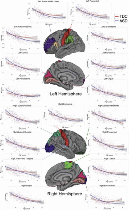
B.A. Zielinski, M.B.D. Prigge, J.A. Nielsen, A.L. Froehlich, T.J. Abildskov, J.S. Anderson, P.T. Fletcher, K.M. Zygmunt, B.G. Travers, N. Lange, A.L. Alexander, E.D. Bigler, J.E. Lainhart. “Longitudinal changes in cortical thickness in autism and typical development,” In Brain, Vol. 137, No. 6, Edited by Dimitri M. Kullmann, pp. 1799--1812. 2014.
DOI: 10.1093/brain/awu083

ABSTRACT

The natural history of brain growth in autism spectrum disorders remains unclear. Cross-sectional studies have identified regional abnormalities in brain volume and cortical thickness in autism, although substantial discrepancies have been reported. Preliminary longitudinal studies using two time points and small samples have identified specific regional differences in cortical thickness in the disorder. To clarify age-related trajectories of cortical development, we examined longitudinal changes in cortical thickness within a large mixed cross-sectional and longitudinal sample of autistic subjects and age- and gender-matched typically developing controls. Three hundred and forty-five magnetic resonance imaging scans were examined from 97 males with autism (mean age = 16.8 years; range 3-36 years) and 60 males with typical development (mean age = 18 years; range 4-39 years), with an average interscan interval of 2.6 years. FreeSurfer image analysis software was used to parcellate the cortex into 34 regions of interest per hemisphere and to calculate mean cortical thickness for each region. Longitudinal linear mixed effects models were used to further characterize these findings and identify regions with between-group differences in longitudinal age-related trajectories. Using mean age at time of first scan as a reference (15 years), differences were observed in bilateral inferior frontal gyrus, pars opercularis and pars triangularis, right caudal middle frontal and left rostral middle frontal regions, and left frontal pole. However, group differences in cortical thickness varied by developmental stage, and were influenced by IQ. Differences in age-related trajectories emerged in bilateral parietal and occipital regions (postcentral gyrus, cuneus, lingual gyrus, pericalcarine cortex), left frontal regions (pars opercularis, rostral middle frontal and frontal pole), left supramarginal gyrus, and right transverse temporal gyrus, superior parietal lobule, and paracentral, lateral orbitofrontal, and lateral occipital regions. We suggest that abnormal cortical development in autism spectrum disorders undergoes three distinct phases: accelerated expansion in early childhood, accelerated thinning in later childhood and adolescence, and decelerated thinning in early adulthood. Moreover, cortical thickness abnormalities in autism spectrum disorders are region-specific, vary with age, and may remain dynamic well into adulthood.


2013


X. Hao, P.T. Fletcher. “Joint Fractional Segmentation and Multi-Tensor Estimation in Diffusion MRI,” In Proceedings of the International Conference on Information Processing in Medical Imaging (IPMI), Lecture Notes in Computer Science (LNCS), pp. (accepted). 2013.

ABSTRACT

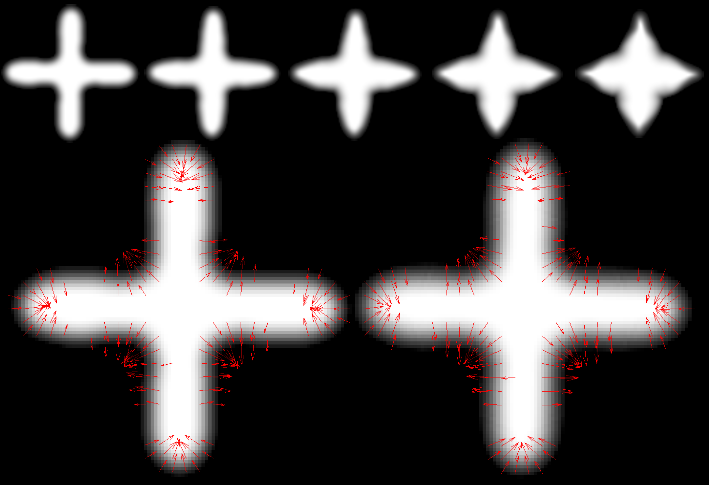
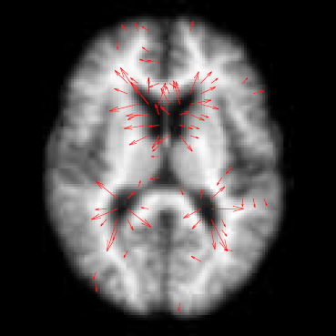
In this paper we present a novel Bayesian approach for fractional segmentation of white matter tracts and simultaneous estimation of a multi-tensor diffusion model. Our model consists of several white matter tracts, each with a corresponding weight and tensor compartment in each voxel. By incorporating a prior that assumes the tensor fields inside each tract are spatially correlated, we are able to reliably estimate multiple tensor compartments in fiber crossing regions, even with low angular diffusion-weighted imaging (DWI). Our model distinguishes the diffusion compartment associated with each tract, which reduces the effects of partial voluming and achieves more reliable statistics of diffusion measurements.We test our method on synthetic data with known ground truth and show that we can recover the correct volume fractions and tensor compartments. We also demonstrate that the proposed method results in improved segmentation and diffusion measurement statistics on real data in the presence of crossing tracts and partial voluming.


N. Sadeghi, M.W. Prastawa, P.T. Fletcher, C. Vachet, Bo Wang, J.H. Gilmore, G. Gerig. “Multivariate Modeling of Longitudinal MRI in Early Brain Development with Confidence Measures,” In Proceedings of the 2013 IEEE 10th International Symposium on Biomedical Imaging (ISBI), pp. 1400--1403. 2013.
DOI: 10.1109/ISBI.2013.6556795

ABSTRACT

The human brain undergoes rapid organization and structuring early in life. Longitudinal imaging enables the study of these changes over a developmental period within individuals through estimation of population growth trajectory and its variability. In this paper, we focus on maturation of white and gray matter as is depicted in structural and diffusion MRI of healthy subjects with repeated scans. We provide a framework for joint analysis of both structural MRI and DTI (Diffusion Tensor Imaging) using multivariate nonlinear mixed effect modeling of temporal changes. Our framework constructs normative growth models for all the modalities that take into account the correlation among the modalities and individuals, along with estimation of the variability of the population trends. We apply our method to study early brain development, and to our knowledge this is the first multimodel longitudinal modeling of diffusion and signal intensity changes for this growth stage. Results show the potential of our framework to study growth trajectories, as well as neurodevelopmental disorders through comparison against the constructed normative models of multimodal 4D MRI.


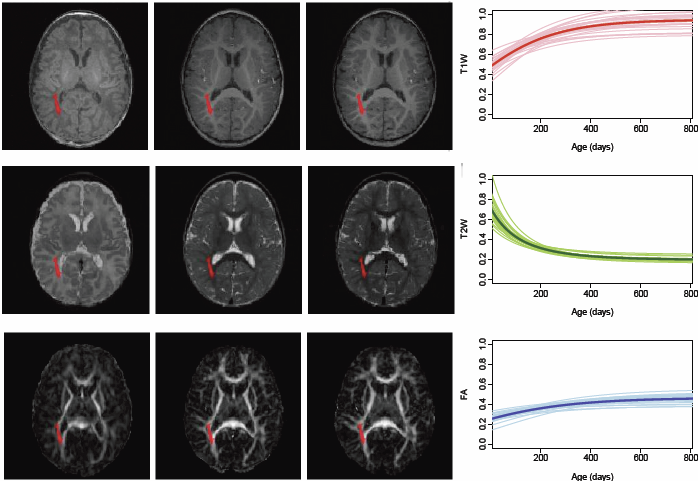
N. Sadeghi, M.W. Prastawa, P.T. Fletcher, J. Wolff, J.H. Gilmore, G. Gerig. 
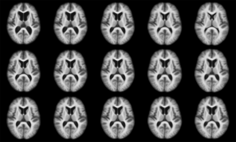
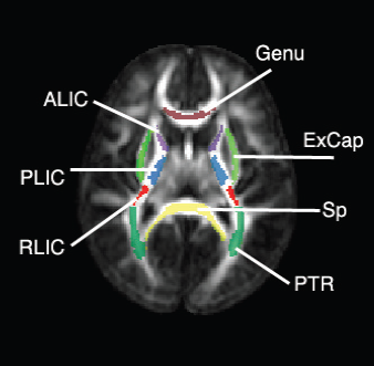
“Regional characterization of longitudinal DT-MRI to study white matter maturation of the early developing brain,” In NeuroImage, Vol. 68, pp. 236--247. March, 2013.
DOI: 10.1016/j.neuroimage.2012.11.040
PubMed ID: 23235270

ABSTRACT

The human brain undergoes rapid and dynamic development early in life. Assessment of brain growth patterns relevant to neurological disorders and disease requires a normative population model of growth and variability in order to evaluate deviation from typical development. In this paper, we focus on maturation of brain white matter as shown in diffusion tensor MRI (DT-MRI), measured by fractional anisotropy (FA), mean diffusivity (MD), as well as axial and radial diffusivities (AD, RD). We present a novel methodology to model temporal changes of white matter diffusion from longitudinal DT-MRI data taken at discrete time points. Our proposed framework combines nonlinear modeling of trajectories of individual subjects, population analysis, and testing for regional differences in growth pattern. We first perform deformable mapping of longitudinal DT-MRI of healthy infants imaged at birth, 1 year, and 2 years of age, into a common unbiased atlas. An existing template of labeled white matter regions is registered to this atlas to define anatomical regions of interest. Diffusivity properties of these regions, presented over time, serve as input to the longitudinal characterization of changes. We use non-linear mixed effect (NLME) modeling where temporal change is described by the Gompertz function. The Gompertz growth function uses intuitive parameters related to delay, rate of change, and expected asymptotic value; all descriptive measures which can answer clinical questions related to quantitative analysis of growth patterns. Results suggest that our proposed framework provides descriptive and quantitative information on growth trajectories that can be interpreted by clinicians using natural language terms that describe growth. Statistical analysis of regional differences between anatomical regions which are known to mature differently demonstrates the potential of the proposed method for quantitative assessment of brain growth and differences thereof. This will eventually lead to a prediction of white matter diffusion properties and associated cognitive development at later stages given imaging data at early stages.


A. Sharma, P.T. Fletcher, J.H. Gilmore, M.L. Escolar, A. Gupta, M. Styner, G. Gerig. “Spatiotemporal Modeling of Discrete-Time Distribution-Valued Data Applied to DTI Tract Evolution in Infant Neurodevelopment,” In Proceedings of the 2013 IEEE 10th International Symposium on Biomedical Imaging (ISBI), pp. 684--687. 2013.
DOI: 10.1109/ISBI.2013.6556567

ABSTRACT

This paper proposes a novel method that extends spatiotemporal growth modeling to distribution-valued data. The method relaxes assumptions on the underlying noise models by considering the data to be represented by the complete probability distributions rather than a representative, single-valued summary statistics like the mean. When summarizing by the latter method, information on the underlying variability of data is lost early in the process and is not available at later stages of statistical analysis. The concept of ’distance’ between distributions and an ’average’ of distributions is employed. The framework quantifies growth trajectories for individuals and populations in terms of the complete data variability estimated along time and space. Concept is demonstrated in the context of our driving application which is modeling of age-related changes along white matter tracts in early neurodevelopment. Results are shown for a single subject with Krabbe's disease in comparison with a normative trend estimated from 15 healthy controls.


N.P. Singh, J. Hinkle, S. Joshi, P.T. Fletcher. “A Vector Momenta Formulation of Diffeomorphisms for Improved Geodesic Regression and Atlas Construction,” In Proceedings of the 2013 IEEE 10th International Symposium on Biomedical Imaging (ISBI), Note: Received Best Student Paper Award, pp. 1219--1222. 2013.
DOI: 10.1109/ISBI.2013.6556700

ABSTRACT

This paper presents a novel approach for diffeomorphic image regression and atlas estimation that results in improved convergence and numerical stability. We use a vector momenta representation of a diffeomorphism's initial conditions instead of the standard scalar momentum that is typically used. The corresponding variational problem results in a closed form update for template estimation in both the geodesic regression and atlas estimation problems. While we show that the theoretical optimal solution is equivalent to the scalar momenta case, the simplification of the optimization problem leads to more stable and efficient estimation in practice. We demonstrate the effectiveness of our method for atlas estimation and geodesic regression using synthetically generated shapes and 3D MRI brain scans.

Keywords: LDDMM, Geodesic regression, Atlas, Vector Momentum


N.P. Singh, J. Hinkle, S. Joshi, P.T. Fletcher. “A Hierarchical Geodesic Model for Diffeomorphic Longitudinal Shape Analysis,” In Proceedings of the International Conference on Information Processing in Medical Imaging (IPMI), Lecture Notes in Computer Science (LNCS), pp. (accepted). 2013.

ABSTRACT

Hierarchical linear models (HLMs) are a standard approach for analyzing data where individuals are measured repeatedly over time. However, such models are only applicable to longitudinal studies of Euclidean data. In this paper, we propose a novel hierarchical geodesic model (HGM), which generalizes HLMs to the manifold setting. Our proposed model explains the longitudinal trends in shapes represented as elements of the group of diffeomorphisms. The individual level geodesics represent the trajectory of shape changes within individuals. The group level geodesic represents the average trajectory of shape changes for the population. We derive the solution of HGMs on diffeomorphisms to estimate individual level geodesics, the group geodesic, and the residual geodesics. We demonstrate the effectiveness of HGMs for longitudinal analysis of synthetically generated shapes and 3D MRI brain scans.


E. Wong, S.P. Awate, P.T. Fletcher. “Adaptive Sparsity in Gaussian Graphical Models,” In Proceedings of the 30th International Conference on Machine Learning (ICML), pp. (accepted). 2013.

ABSTRACT

An effective approach to structure learning and parameter estimation for Gaussian graphical models is to impose a sparsity prior, such as a Laplace prior, on the entries of the precision matrix. Such an approach involves a hyperparameter that must be tuned to control the amount of sparsity. In this paper, we introduce a parameter-free method for estimating a precision matrix with sparsity that adapts to the data automatically. We achieve this by formulating a hierarchical Bayesian model of the precision matrix with a noninformative Jeffreys' hyperprior. We also naturally enforce the symmetry and positivede definiteness constraints on the precision matrix by parameterizing it with the Cholesky decomposition. Experiments on simulated and real (cell signaling) data demonstrate that the proposed approach not only automatically adapts the sparsity of the model, but it also results in improved estimates of the precision matrix compared to the Laplace prior model with sparsity parameter chosen by cross-validation.


M. Zhang, N.P. Singh, P.T. Fletcher. “Bayesian Estimation of Regularization and Atlas Building in Diffeomorphic Image Registration,” In Proceedings of the International Conference on Information Processing in Medical Imaging (IPMI), Lecture Notes in Computer Science (LNCS), pp. (accepted). 2013.

ABSTRACT

This paper presents a generative Bayesian model for diffeomorphic image registration and atlas building. We develop an atlas estimation procedure that simultaneously estimates the parameters controlling the smoothness of the diffeomorphic transformations. To achieve this, we introduce a Monte Carlo Expectation Maximization algorithm, where the expectation step is approximated via Hamiltonian Monte Carlo sampling on the manifold of diffeomorphisms. An added benefit of this stochastic approach is that it can successfully solve difficult registration problems involving large deformations, where direct geodesic optimization fails. Using synthetic data generated from the forward model with known parameters, we demonstrate the ability of our model to successfully recover the atlas and regularization parameters. We also demonstrate the effectiveness of the proposed method in the atlas estimation problem for 3D brain images.


M. Zhang, P.T. Fletcher. “Probabilistic Principal Geodesic Analysis,” In Proceedings of the 2013 Conference on Neural Information Processing Systems (NIPS), pp. (accepted). 2013.

ABSTRACT

Principal geodesic analysis (PGA) is a generalization of principal component analysis (PCA) for dimensionality reduction of data on a Riemannian manifold. Currently PGA is defined as a geometric fit to the data, rather than as a probabilistic model. Inspired by probabilistic PCA, we present a latent variable model for PGA that provides a probabilistic framework for factor analysis on manifolds. To compute maximum likelihood estimates of the parameters in our model, we develop a Monte Carlo Expectation Maximization algorithm, where the expectation is approximated by Hamiltonian Monte Carlo sampling of the latent variables. We demonstrate the ability of our method to recover the ground truth parameters in simulated sphere data, as well as its effectiveness in analyzing shape variability of a corpus callosum data set from human brain images.


X. Zhu, Y. Gur, W. Wang, P.T. Fletcher. “Model Selection and Estimation of Multi-Compartment Models in Diffusion MRI with a Rician Noise Model,” In Proceedings of the International Conference on Information Processing in Medical Imaging (IPMI), Lecture Notes in Computer Science (LNCS), Vol. 23, pp. 644--655. 2013.
PubMed ID: 24684006

ABSTRACT

Multi-compartment models in diffusion MRI (dMRI) are used to describe complex white matter fiber architecture of the brain. In this paper, we propose a novel multi-compartment estimation method based on the ball-and-stick model, which is composed of an isotropic diffusion compartment (\"ball\") as well as one or more perfectly linear diffusion compartments (\"sticks\"). To model the noise distribution intrinsic to dMRI measurements, we introduce a Rician likelihood term and estimate the model parameters by means of an Expectation Maximization (EM) algorithm. This paper also addresses the problem of selecting the number of fiber compartments that best fit the data, by introducing a sparsity prior on the volume mixing fractions. This term provides automatic model selection and enables us to discriminate different fiber populations. When applied to simulated data, our method provides accurate estimates of the fiber orientations, diffusivities, and number of compartments, even at low SNR, and outperforms similar methods that rely on a Gaussian noise distribution assumption. We also apply our method to in vivo brain data and show that it can successfully capture complex fiber structures that match the known anatomy.


2012


M. Datar, P. Muralidharan, A. Kumar, S. Gouttard, J. Piven, G. Gerig, R.T. Whitaker, P.T. Fletcher. “Mixed-Effects Shape Models for Estimating Longitudinal Changes in Anatomy,” In Spatio-temporal Image Analysis for Longitudinal and Time-Series Image Data, Lecture Notes in Computer Science, Vol. 7570, Springer Berlin / Heidelberg, pp. 76--87. 2012.
ISBN: 978-3-642-33554-9
DOI: 10.1007/978-3-642-33555-6_7

ABSTRACT

In this paper, we propose a new method for longitudinal shape analysis that ts a linear mixed-e ects model, while simultaneously optimizing correspondences on a set of anatomical shapes. Shape changes are modeled in a hierarchical fashion, with the global population trend as a xed e ect and individual trends as random e ects. The statistical signi cance of the estimated trends are evaluated using speci cally designed permutation tests. We also develop a permutation test based on the Hotelling T2 statistic to compare the average shapes trends between two populations. We demonstrate the bene ts of our method on a synthetic example of longitudinal tori and data from a developmental neuroimaging study.

Keywords: Computer Science


J. Hinkle, P. Muralidharan, P.T. Fletcher, S. Joshi. “Polynomial Regression on Riemannian Manifolds,” In arXiv, Vol. 1201.2395, 2012.

ABSTRACT

In this paper we develop the theory of parametric polynomial regression in Riemannian manifolds and Lie groups. We show application of Riemannian polynomial regression to shape analysis in Kendall shape space. Results are presented, showing the power of polynomial regression on the classic rat skull growth data of Bookstein as well as the analysis of the shape changes associated with aging of the corpus callosum from the OASIS Alzheimer's study.


W. Liu, S. Awate, P.T. Fletcher. “Group Analysis of Resting-State fMRI by Hierarchical Markov Random Fields,” In Medical Image Computing and Computer-Assisted Intervention – MICCAI 2012, Lecture Notes in Computer Science (LNCS), Vol. 7512, pp. 189--196. 2012.
ISBN: 978-3-642-33453-5
DOI: 10.1007/978-3-642-33454-2_24

ABSTRACT

Identifying functional networks from resting-state functional MRI is a challenging task, especially for multiple subjects. Most current studies estimate the networks in a sequential approach, i.e., they identify each individual subject's network independently to other subjects, and then estimate the group network from the subjects networks. This one-way flow of information prevents one subject's network estimation benefiting from other subjects. We propose a hierarchical Markov Random Field model, which takes into account both the within-subject spatial coherence and between-subject consistency of the network label map. Both population and subject network maps are estimated simultaneously using a Gibbs sampling approach in a Monte Carlo Expectation Maximization framework. We compare our approach to two alternative groupwise fMRI clustering methods, based on K-means and Normalized Cuts, using both synthetic and real fMRI data.We show that our method is able to estimate more consistent subject label maps, as well as a stable group label map.


P. Muralidharan, P.T. Fletcher. “Sasaki Metrics for Analysis of Longitudinal Data on Manifolds,” In Proceedings of the 2012 IEEE conference on Computer Vision and Pattern Recognition (CVPR), pp. 1027--1034. 2012.
DOI: 10.1109/CVPR.2012.6247780

ABSTRACT

Longitudinal data arises in many applications in which the goal is to understand changes in individual entities over time. In this paper, we present a method for analyzing longitudinal data that take values in a Riemannian manifold. A driving application is to characterize anatomical shape changes and to distinguish between trends in anatomy that are healthy versus those that are due to disease. We present a generative hierarchical model in which each individual is modeled by a geodesic trend, which in turn is considered as a perturbation of the mean geodesic trend for the population. Each geodesic in the model can be uniquely parameterized by a starting point and velocity, i.e., a point in the tangent bundle. Comparison between these parameters is achieved through the Sasaki metric, which provides a natural distance metric on the tangent bundle. We develop a statistical hypothesis test for differences between two groups of longitudinal data by generalizing the Hotelling T2 statistic to manifolds. We demonstrate the ability of these methods to distinguish differences in shape changes in a comparison of longitudinal corpus callosum data in subjects with dementia versus healthily aging controls.


N. Sadeghi, M.W. Prastawa, P.T. Fletcher, J.H. Gilmore, W. Lin, G. Gerig. “Statistical Growth Modeling of Longitudinal DT-MRI for Regional Characterization of Early Brain Development,” In Proceedings of IEEE ISBI 2012, pp. 1507--1510. 2012.
DOI: 10.1109/ISBI.2012.6235858

ABSTRACT

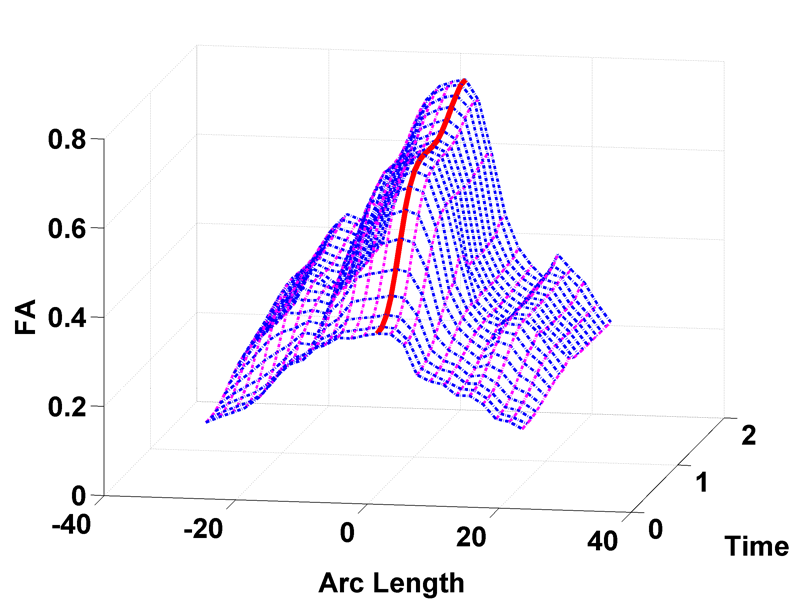
A population growth model that represents the growth trajectories of individual subjects is critical to study and understand neurodevelopment. This paper presents a framework for jointly estimating and modeling individual and population growth trajectories, and determining significant regional differences in growth pattern characteristics applied to longitudinal neuroimaging data. We use non-linear mixed effect modeling where temporal change is modeled by the Gompertz function. The Gompertz function uses intuitive parameters related to delay, rate of change, and expected asymptotic value; all descriptive measures which can answer clinical questions related to growth. Our proposed framework combines nonlinear modeling of individual trajectories, population analysis, and testing for regional differences. We apply this framework to the study of early maturation in white matter regions as measured with diffusion tensor imaging (DTI). Regional differences between anatomical regions of interest that are known to mature differently are analyzed and quantified. Experiments with image data from a large ongoing clinical study show that our framework provides descriptive, quantitative information on growth trajectories that can be directly interpreted by clinicians. To our knowledge, this is the first longitudinal analysis of growth functions to explain the trajectory of early brain maturation as it is represented in DTI.


N.P. Singh, A.Y. Wang, P. Sankaranarayanan, P.T. Fletcher, S. Joshi. “Genetic, Structural and Functional Imaging Biomarkers for Early Detection of Conversion from MCI to AD,” In Proceedings of Medical Image Computing and Computer-Assisted Intervention MICCAI 2012, Vol. 7510, pp. 132--140. 2012.
DOI: 10.1007/978-3-642-33415-3_17

ABSTRACT

With the advent of advanced imaging techniques, genotyping, and methods to assess clinical and biological progression, there is a growing need for a unified framework that could exploit information available from multiple sources to aid diagnosis and the identification of early signs of Alzheimer’s disease (AD). We propose a modeling strategy using supervised feature extraction to optimally combine highdimensional imaging modalities with several other low-dimensional disease risk factors. The motivation is to discover new imaging biomarkers and use them in conjunction with other known biomarkers for prognosis of individuals at high risk of developing AD. Our framework also has the ability to assess the relative importance of imaging modalities for predicting AD conversion. We evaluate the proposed methodology on the Alzheimer’s Disease Neuroimaging Initiative (ADNI) database to predict conversion of individuals with Mild Cognitive Impairment (MCI) to AD, only using information available at baseline.

Keywords: adni