Clinical Application of Electrocardiographic Inverse Solution
Petr Stovicek, MD/Ph.D.
Charles University Hospital, Prague, Czech Republic
Background
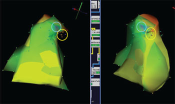 |
| ECG images (potential distributions) obtained by inverse solution computed on the triangularized mesh (model) of the right ventricle of a human subject treated for ventricular arrhythmia. The blue circle surrounds the region of initial QRS negativity with the potential minimum at its center. Overlapping is the CARTO right ventricle map (it is the rougher and light green colored surface with its nodes in the form of small balls). The yellow circle containing orange and red balls marks the earliest activation sites and also sites of the successful ablation on the CARTO |
This collaboration has special importance for two reasons. First, Dr. Stovicek is a clinical electrophysiologist who downloaded, installed, and learned to use SCIRun completely on his own and then reported his progress to us only when he had questions about relative subtleties of the regularization approaches implements in SCIRun. We have noted a growing ease with which users can access, install, and use the Center software, especially since we achieved binary downloads for the major platforms (Windows and OSX) and Dr. Stovicek's success was a marvelous illustration of the impact. The second notable feature of this emerging collaboration is the Dr. Stovicek is a practicing clinical cardiac electrophysiologist at a leading clinic in his home country and believes that computational inverse solutions are reaching a state of utility that could affect his practice. He has published a short report on a case in which the predicted location of an early activation site was predicted using SCIRun and a generic torso model1 and is now excited to continue to refine and experiment with this approach. He has also applied for grant funding to expand the scope of the project to include a leading Cardiology group in Newcastle, Great Britain, where he and his colleagues would apply the inverse approach to a wider set of patients than he currently has available, and would be able to create patient specific geometric models with which to explore the clinical application of this method.
Aims
The results in the figure are very promising indications that it is possible to apply electrocardiographic inverse imaging to clinical cases and the goals of the collaboration are to expand and explore this opportunity. Carrying our this project in such close communication with a practicing clinician will ensure that all aspects of the resulting algorithms and software are geared toward translation and that close feedback will accelerate progress and improve the likelihood of both technical success and clinical impact. The resulting specific aims of this collaboration include the following:
- Assist collaborator to create patient specific geometries for use with inverse solution.
- Assist collaborator with mapping of body surface potentials to torso geometry to provide simulation boundary conditions.
- Explore opportunities to apply activation time based source model in order to capture epicardial and endocardial activation times from inverse solution.
- Assist collaborator with exploration of regularization techniques to create robust and clinically useful solutions.
Publications
P. Stovicek, S. Havranek, J. Simek, M. Zbornik, M. Micek, and O. Kittnar. "Isopotential ECG Imaging Correctly Identified Endocardial Ectopic Activation Site in the Case of Arrhythmia from Right Ventricular Outflow Tract". In World Congress on Med. Phys. and Biomed. Eng., volume 25/IV, pages 1965–1968, Heidelberg, 2009. Springer.