|
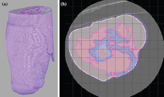
| Figure from B.M. Issacson, et al., A unilateral hierarchical model was assembled as a representative image consisting of skin (purple) adipose tissue (yellow), musculature (pink), bone (blue), bone marrow (orange), and internal organs (green) (a). Each tissue type was assigned a specific conductivity using SCIRun. A large serpentine-like mass of HO was identified in the medial aspect of the residual limb, and was demonstrated in more detail in an axial cross section of the affected limb (b). |
Osseointegration is a surgical procedure that provides direct skeletal attachment between an implant and host tissue with proven success in dental, auricle, and transfemoral implants. However, one challenge with using natural biological fixation is attaining a strong skeletal interlock at the implant interface, a prerequisite for long-term implant function. Utilizing metallic implants as a means of biological fixation has been the objective of orthopedic surgeons over the past two centuries. However, controlling osteogenesis at the implant interface, which is essential for providing strong skeletal fixation, remains challenging. Regulated electrical stimulation has proven effective in fracture healing and non-traumatized bone models, but has not been investigated in a percutaneous osseointegrated implant system. One advantage of the veteran patient population is that an orthopedic implant protrudes from the residual limb functioning as an exoprosthesis attachment and may operate as a potential cathode for an external electrical stimulation device.